Sections of forearm and hand
Transverse section of right wrist
Stanford holds the copyright to the David L. Bassett anatomical images and has assigned
Creative Commons license Attribution-Share
Alike 4.0 International to all of the images.
For additional information regarding use and permissions,
please contact the Medical History Center.
Image #112-2
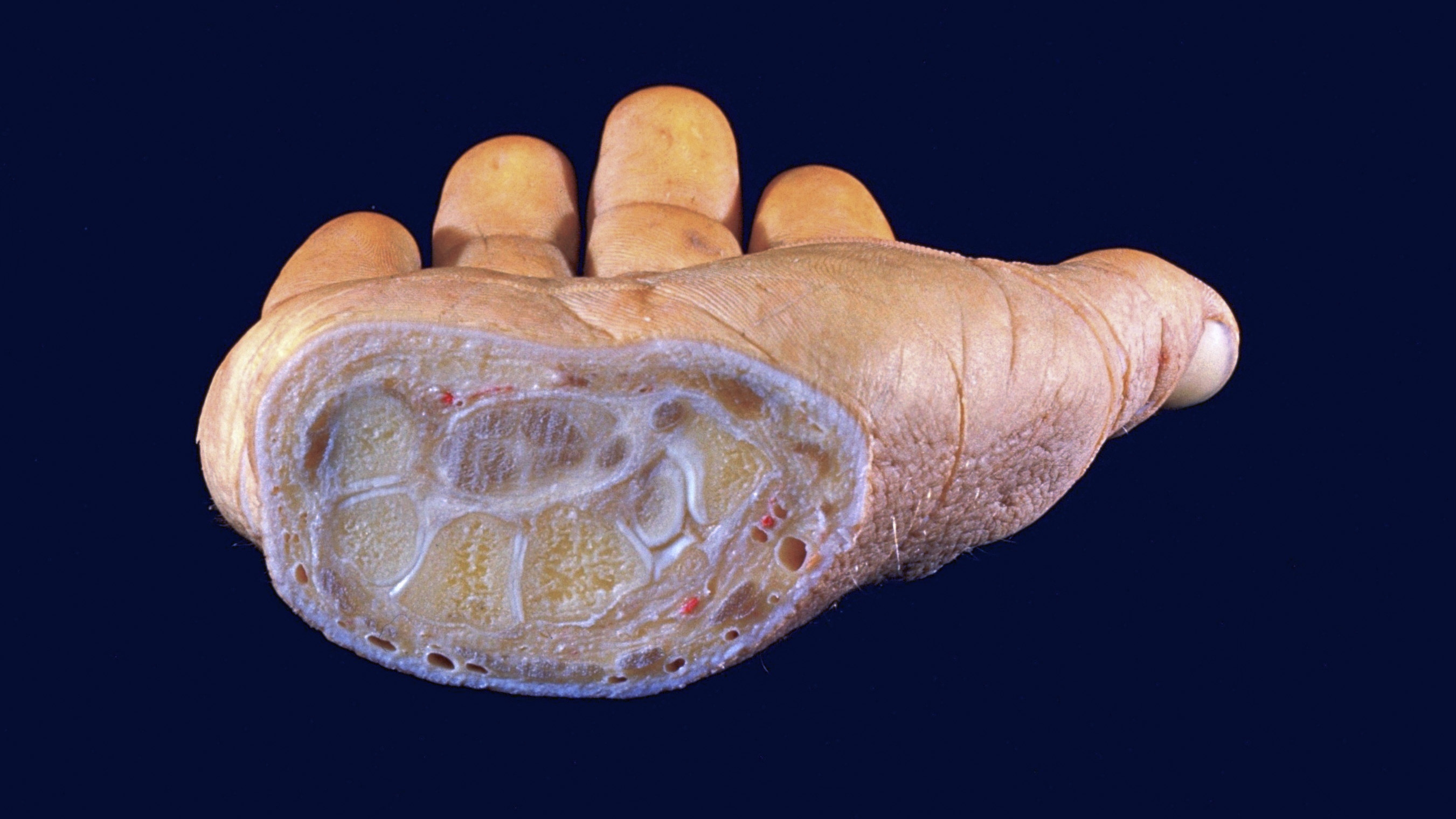
Sections of forearm and hand
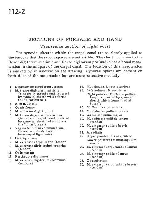
Transverse section of right wrist
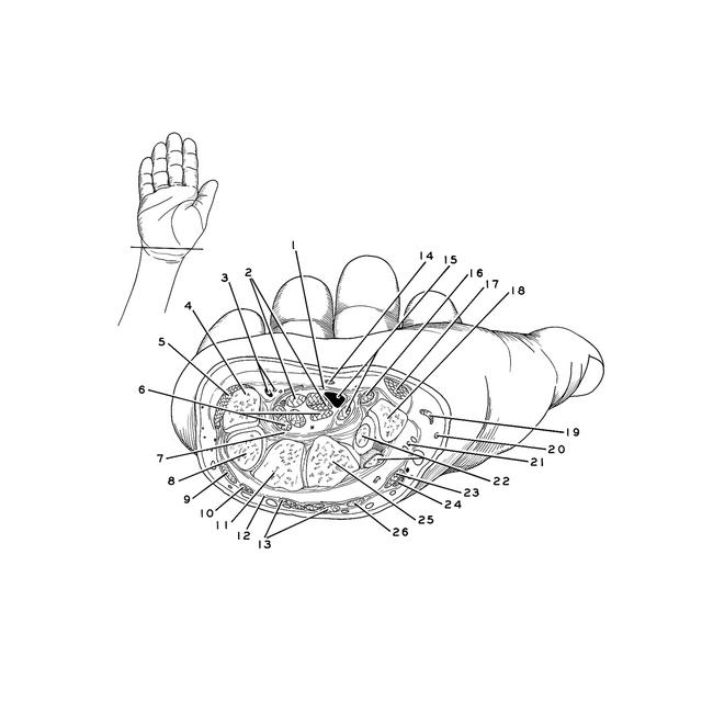
The synovial sheaths within the carpal canal are so closely applied to the tendons that the serous spaces are not visible. The sheath common to the flexor digitorum sublimis and flexor digitorum profundus has a broad mesotendon in the midpart of the carpal canal. The location of this mesotendon is marked by an asterisk on the drawing. Synovial spaces are present on both sides of the mesotendon but are more extensive medially.
- Transverse carpal ligament
- Flexor digitorum superficialis (tendons in carpal canal, invested by synovial sheath which forms the "ulnar bursa")
- Ulnar artery and nerve
- Pisiform bone
- Abductor digiti minimi muscle
- Flexor digitorum profundus muscle (tendons in carpal canal, invested by synovial sheath which forms the "ulnar bursa")
- Sheath of common tendon of flexor muscles (blended with intercarpal ligaments)
- Triquetral bone
- Extensor carpi ulnaris muscle (tendon)
- Extensor digiti minimi muscle (tendon)
- Hamate bone
- Dorsal fascia of the hand
- Common extensor digitorum muscle (tendons)
- Palmaris longus muscle (tendon)
- Left pointer: Median nerve Right pointer: Flexor pollicis longus muscle (invested by synovial sheath which forms "radial bursa")
- Flexor carpi radialis muscle
- Abductor pollicis brevis muscle
- Trapezium bone
- Abductor pollicis longus muscle (tendon)
- Extensor pollicis brevis muscle (tendon)
- Radial artery
- Upper pointer: Scaphoid bone Lower pointer: Trapezoid bone
- Extensor carpi radialis longus muscle (tendon)
- Extensor pollicis longus muscle (tendon)
- Capitate bone
- Extensor carpi radialis brevis muscle (tendon)