Dorsal aspect of hand
Compartments for extensor tendons deep to dorsal carpal ligament, lateral view of right hand
Stanford holds the copyright to the David L. Bassett anatomical images and has assigned
Creative Commons license Attribution-Share
Alike 4.0 International to all of the images.
For additional information regarding use and permissions,
please contact the Medical History Center.
Image #109-7
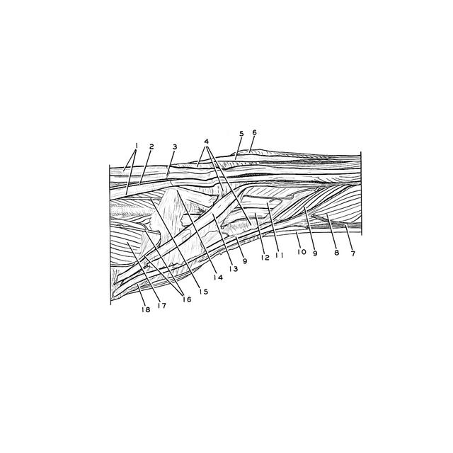
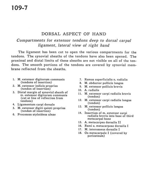
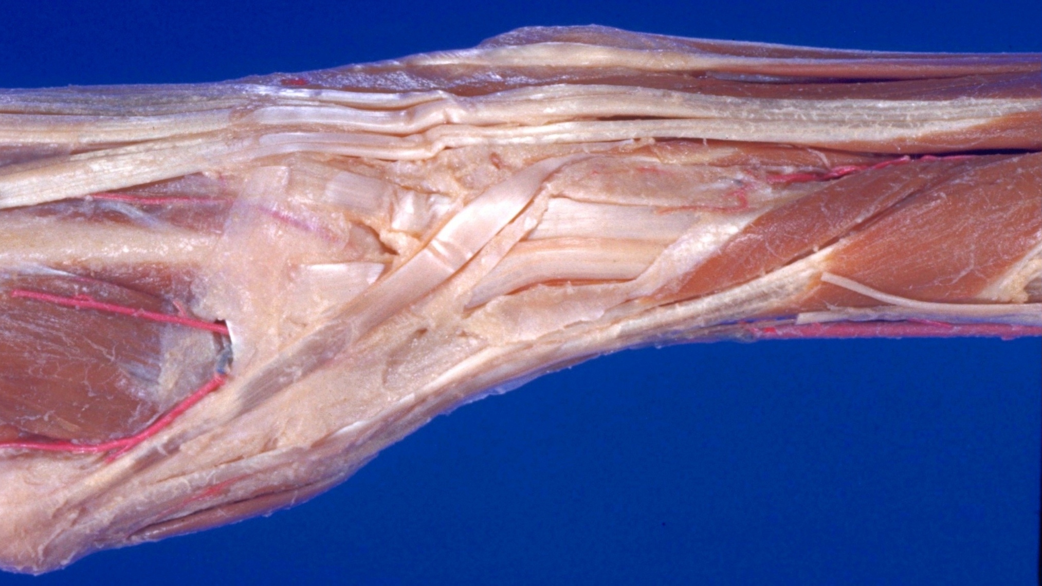
Dorsal aspect of hand
Compartments for extensor tendons deep to dorsal carpal ligament, lateral view of right hand
The ligament has been cut to open the various compartments for the tendons. The synovial sheaths of the tendons have also been opened. The proximal and distal limits of these sheaths are not visible on all of the tendons. The smooth portions of the tendons are covered by synovial membrane reflected from the sheaths.
- Common extensor digitorum muscle (tendons of insertion)
- Extensor indicis muscle (tendon of insertion)
- Distal margin of synovial sheath of common extensor digitorum muscle (cut at line of reflection from tendons)
- Dorsal carpal ligament
- Extensor digiti minimi muscle (tendon of insertion)
- Styloid process of ulna
- Superficial branch of radial nerve
- Abductor pollicis longus muscle
- Extensor pollicis brevis muscle
- Radial artery
- Extensor carpi radialis brevis muscle (tendon)
- Extensor carpi radialis longus muscle (tendon)
- Extensor pollicis longus muscle (tendon)
- Insertion of extensor carpi radialis brevis muscle into base of third metacarpal bone
- Dorsal metacarpal artery II
- Branches of dorsal metacarpal artery I
- Dorsal interosseous muscle I
- Metacarpal I (covered by periosteum)