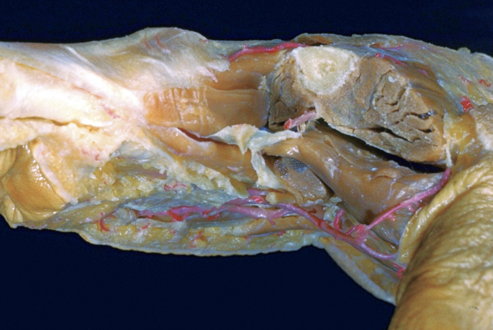
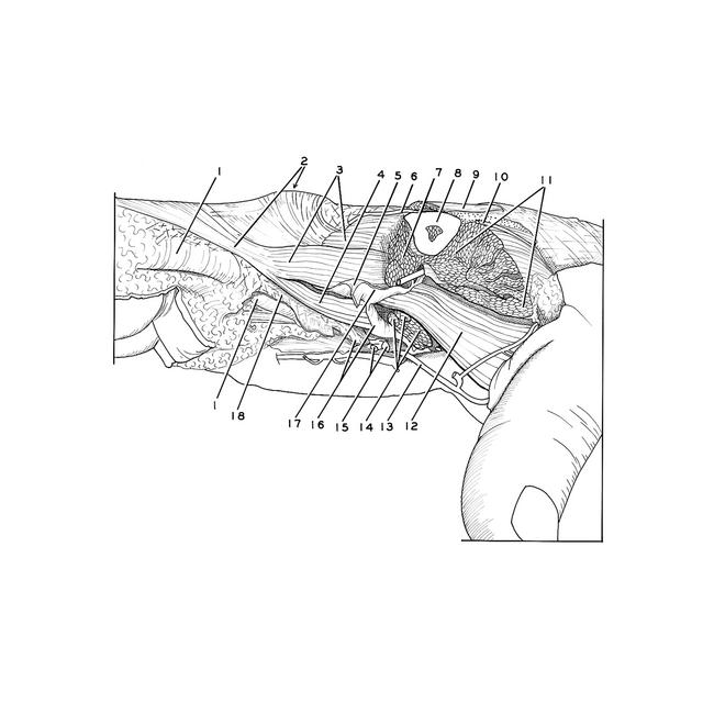
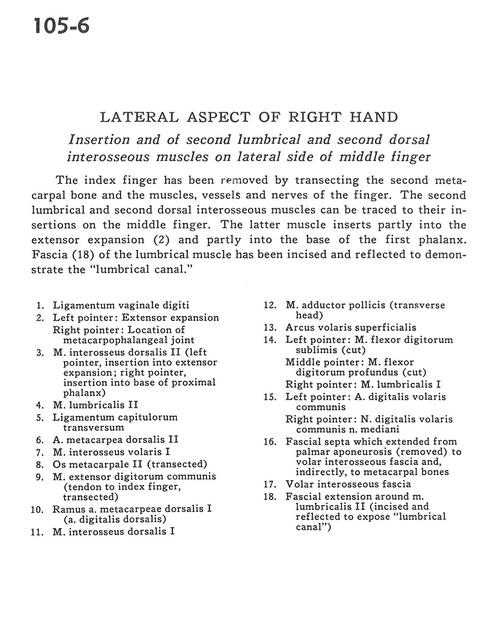
Lateral aspect of right hand
Insertion and of second lumbrical and second dorsal interosseous muscles on lateral side of middle finger
Stanford holds the copyright to the David L. Bassett anatomical images and has assigned
Creative Commons license Attribution-Share
Alike 4.0 International to all of the images.
For additional information regarding use and permissions,
please contact the Medical History Center.
Image #105-6
Lateral aspect of right hand
Insertion and of second lumbrical and second dorsal interosseous muscles on lateral side of middle finger
The index finger has been removed by transecting the second metacarpal bone and the muscles, vessels and nerves of the finger. The second lumbrical and second dorsal interosseous muscles can be traced to their insertions on the middle finger. The latter muscle inserts partly into the extensor expansion (2) and partly into the base of the first phalanx. Fascia (18) of the lumbrical muscle has been incised and reflected to demonstrate the "lumbrical canal."
- Ligament of digital sheath
- Left pointer: Extensor expansion Right pointer: Location of metacarpophalangeal joint
- Dorsal interosseous muscle II (left pointer, insertion into extensor expansion; right pointer, insertion into base of proximal phalanx)
- Lumbrical muscle II
- Deep transverse metacarpal ligament
- Dorsal metacarpal artery II
- Anterior interosseous muscle I
- Metacarpal II (transected)
- Common extensor digitorum muscle (tendon to index finger, transected)
- Branch dorsal metacarpal artery I (dorsal digital artery)
- Dorsal interosseous muscle I
- Adductor pollicis muscle (transverse head)
- Superficial palmar arch
- Left pointer: Flexor digitorum superficialis (cut) Middle pointer: Flexor digitorum profundus muscle (cut) Right pointer: Lumbrical muscle I
- Left pointer: Anterior common digital artery Right pointer: Common palmar digital nerve of median nerve
- Fascial septa which extended from palmar aponeurosis (removed) to anterior interosseous fascia and, indirectly, to metacarpal bones
- Anterior interosseous fascia
- Fascial extension around lumbrical muscle II (incised and reflected to expose "lumbrical canal")