Volar aspect of right hand
Flexor tendons reflected from carpal canal; volar interosseous fascia
Stanford holds the copyright to the David L. Bassett anatomical images and has assigned
Creative Commons license Attribution-Share
Alike 4.0 International to all of the images.
For additional information regarding use and permissions,
please contact the Medical History Center.
Image #104-1
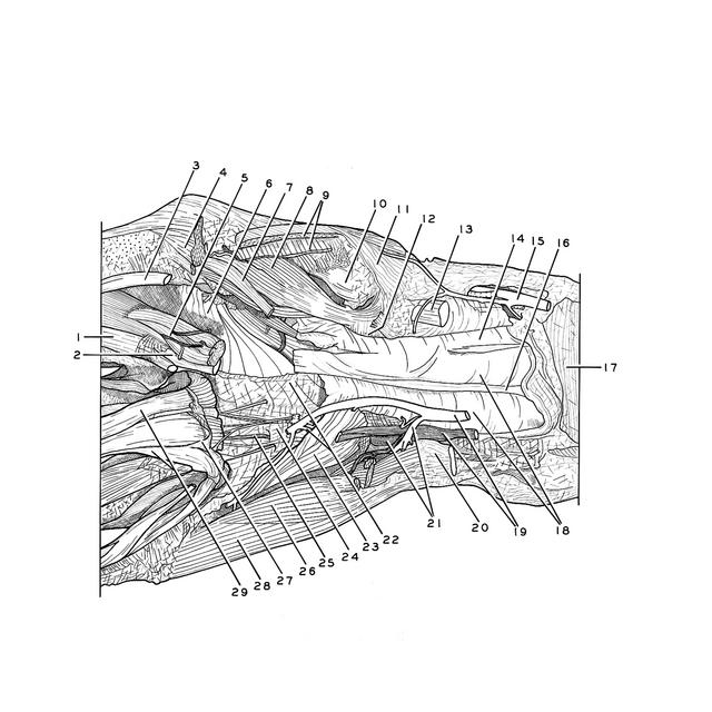
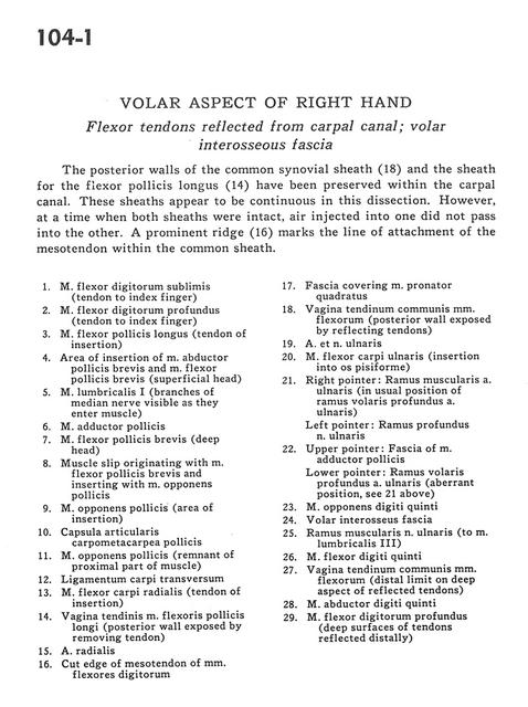
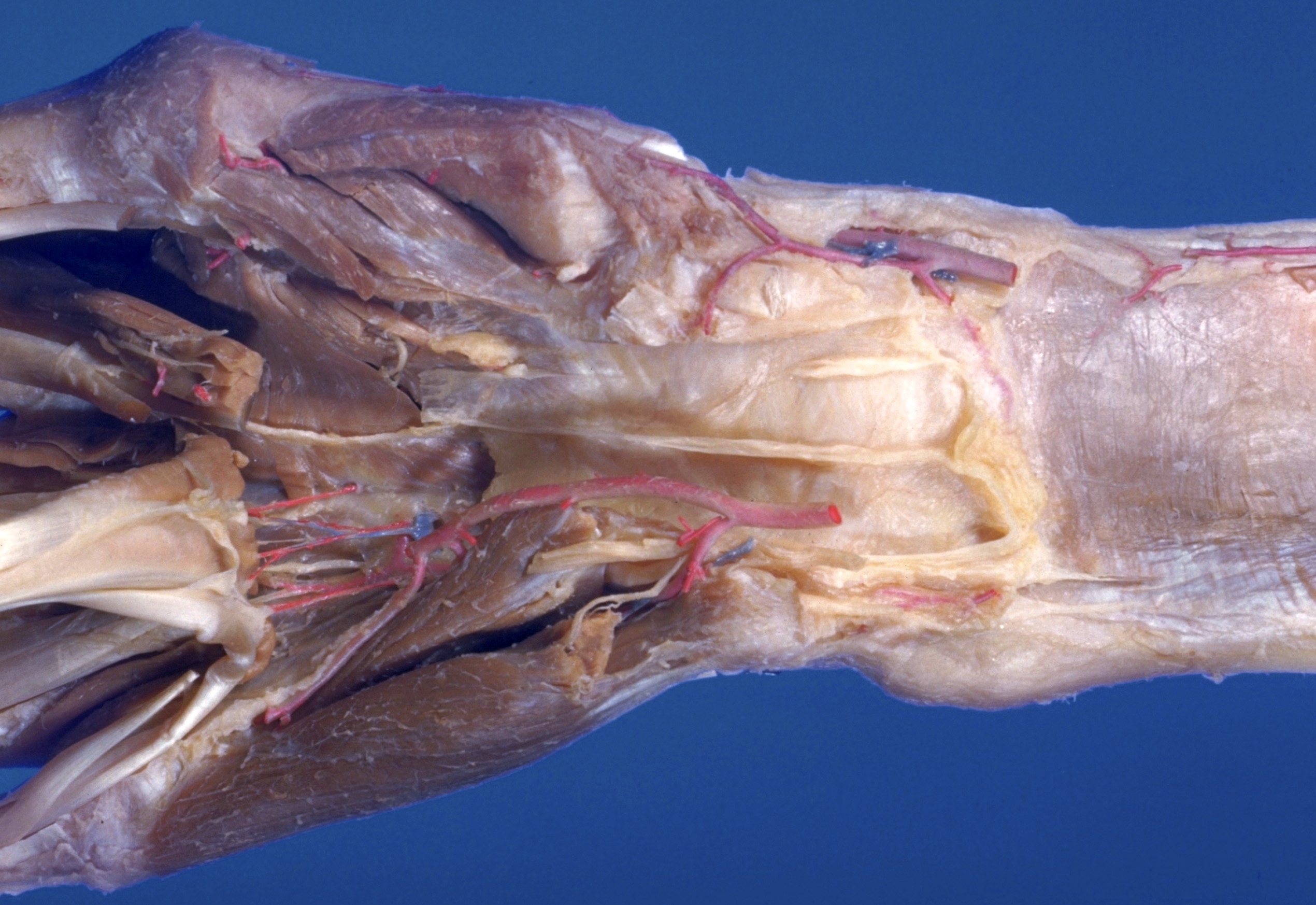
Volar aspect of right hand
Flexor tendons reflected from carpal canal; volar interosseous fascia
The posterior walls of the common synovial sheath (18) and the sheath of the flexor pollicis longus (14) have been preserved within the carpal canal. These sheaths appear to be continuous in this dissection. However, at a time when both sheaths were intact, air injected into one did not pass into the other. A prominent ridge (16) marks the line of attachment of the mesotendon within the common sheath.
- Flexor digitorum superficialis (tendon to index finger)
- Flexor digitorum profundus muscle (tendon to index finger)
- Flexor pollicis longus muscle (tendon of insertion)
- Area of insertion of abductor pollicis brevis muscle and flexor pollicis brevis muscle (superficial head)
- Lumbrical muscle I (branches of median nerve visible as they enter muscle)
- Adductor pollicis muscle
- Flexor pollicis brevis muscle (deep head)
- Muscle slip originating with flexor pollicis brevis muscle and inserting with opponens pollicis muscle
- Opponens pollicis muscle (area of insertion)
- Carpometacarpal joint capsule
- Opponens pollicis muscle (remnant of proximal part of muscle)
- Transverse carpal ligament
- Flexor carpi radialis muscle (tendon of insertion)
- Tendon sheath of flexor pollicis longus muscle (posterior wall exposed by removing tendon)
- Radial artery
- Cut edge of mesotendon of flexor digitorum muscles
- Fascia covering pronator quadratus muscle
- Sheath of common tendon of flexor muscles (posterior wall exposed by reflecting tendons)
- Ulnar artery and nerve
- Flexor carpi ulnaris muscle (insertion into pisiform bone)
- Right pointer: Muscular branch ulnar artery (in usual position of deep anterior branch ulnar artery) Left pointer: Deep branch of ulnar nerve
- Upper pointer: Fascia of adductor pollicis muscle Lower pointer: Deep anterior branch ulnar artery (aberrant position, see 21 above)
- Opponens digiti minimi muscle
- Anterior interosseus fascia
- Muscular branch of ulnar nerve (to lumbrical muscle III)
- Flexor digiti minimi muscle
- Sheath of common tendon of flexor muscles (distal limit on deep aspect of reflected tendons)
- Abductor digiti minimi muscle
- Flexor digitorum profundus muscle (deep surfaces of tendons reflected distally)