Volar aspect of right hand
Synovial sheath in carpus canal for tendon of flexor pollicis longus muscle (radial bursa)
Stanford holds the copyright to the David L. Bassett anatomical images and has assigned
Creative Commons license Attribution-Share
Alike 4.0 International to all of the images.
For additional information regarding use and permissions,
please contact the Medical History Center.
Image #103-1
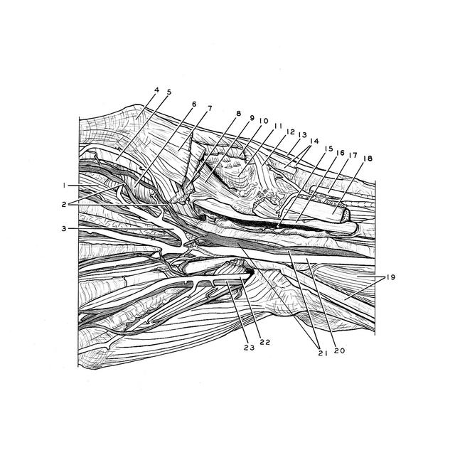
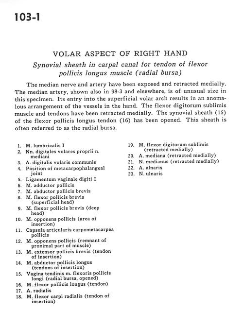
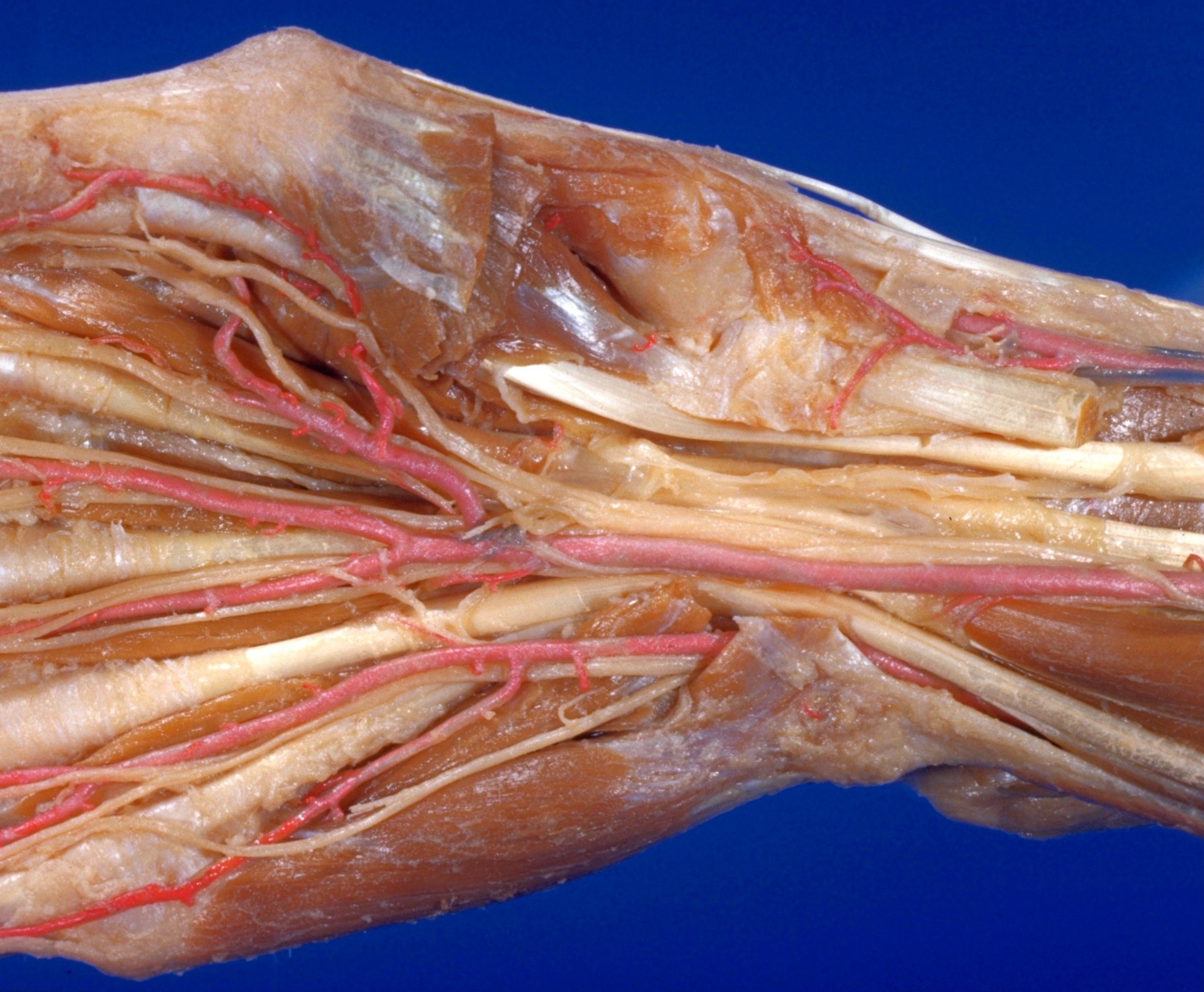
Volar aspect of right hand
Synovial sheath in carpus canal for tendon of flexor pollicis longus muscle (radial bursa)
The median nerve and artery have been exposed and retracted medially. The median artery, shown also in 98-3 and elsewhere, is of unusual size in this specimen. Its entry into the superficial volar arch results in an anomalous arrangement of the vessels in the hand. The flexor digitorum sublimis muscle and tendons have been retracted medially. The synovial sheath (15) of the flexor pollicis longus tendon (16) has been opened. This sheath is often referred to as the radial bursa.
- Lumbrical muscle I
- Proper palmar digital nerves of median nerve
- Anterior common digital artery
- Position of metacarpophalangeal joint
- Ligament of digital sheath
- Adductor pollicis muscle
- Abductor pollicis brevis muscle
- Flexor pollicis brevis muscle (superficial head)
- Flexor pollicis brevis muscle (deep head)
- Opponens pollicis muscle (area of insertion)
- Metacarpotrapezial joint capsule
- Opponens pollicis muscle (remnant of proximal part of muscle)
- Extensor pollicis brevis (tendon of insertion)
- Abductor pollicis longus muscle (tendons of insertion)
- Tendon sheath of flexoris pollicis longus muscle (radial bursa, opened)
- Flexor pollicis longus muscle (tendon)
- Radial artery
- Flexor carpi radialis muscle (tendon of insertion)
- Flexor digitorum superficialis (retracted medially)
- Median artery (retracted medially)
- Median nerve (retracted medially)
- Ulnar artery
- Ulnar nerve