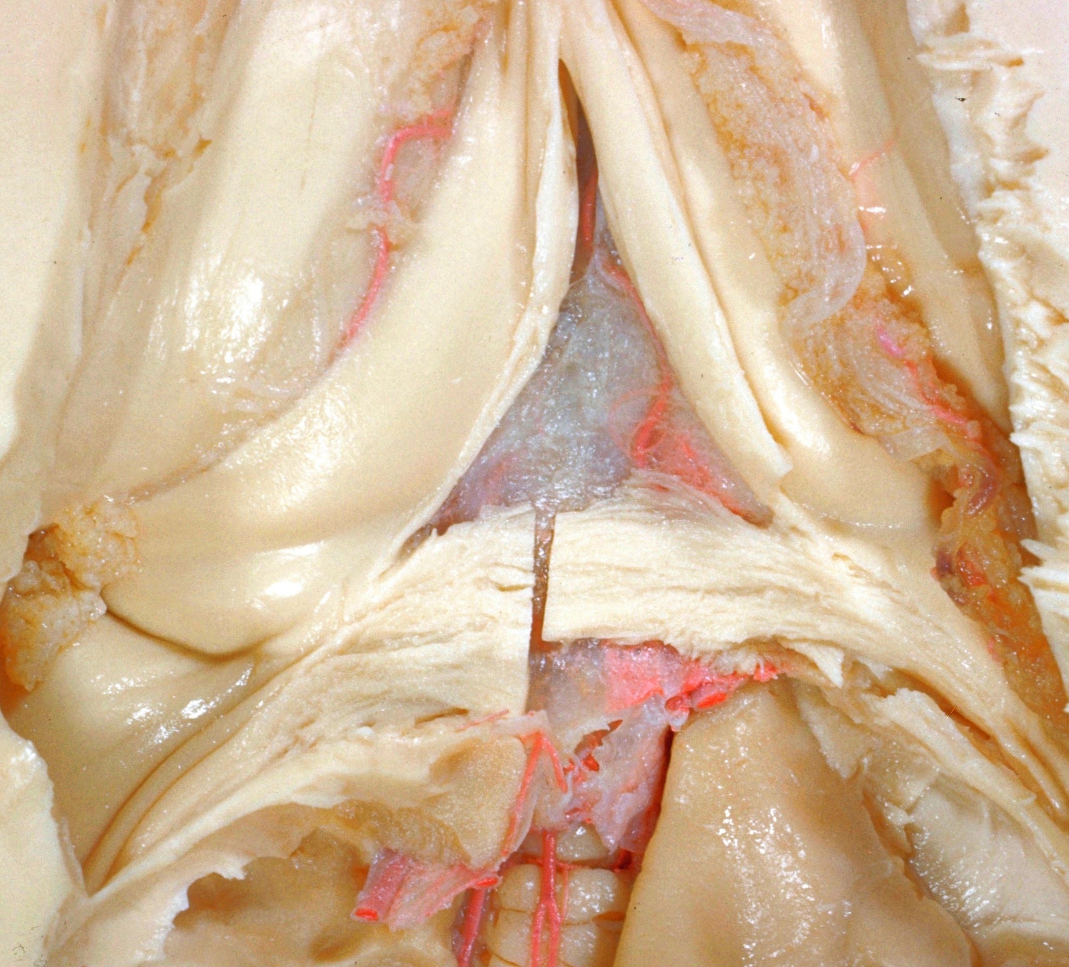
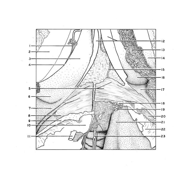
Exploration of the brain from its superior aspect
Hippocampal commissure, crura of fornix and transverse fissure
Stanford holds the copyright to the David L. Bassett anatomical images and has assigned
Creative Commons license Attribution-Share
Alike 4.0 International to all of the images.
For additional information regarding use and permissions,
please contact the Medical History Center.
Image #10-4
Exploration of the brain from its superior aspect
Hippocampal commissure, crura of fornix and transverse fissure
Close-up view of the region of the hippocampal commisure which in this specimen consists of only a few fibers crossing between the hippocampi, posterior to the crura of the fornix. Some of the commissural fibers which are visible to the left of the midline cut belong to the corpus callosum and can be traced into the most medial part of the forceps major. The prominence formed by these fibers within the ventricle is named the bulbus cornu posterioris. To the right of the sagittal cut more callosal fibers have been removed to reveal the meninges and vessels underlying the splenium.
- Choroidal branch of posterior cerebral artery lying along choroidal fissure of lateral ventricle
- Lamina affixa
- Fornix (crus)
- Choroidal fissure
- Hippocampal commissure (cut through in midline)
- Hippocampus
- Calcar avis
- Fibers of occipital part radiations of corpus callosum
- Fibers of the cingulum (cut across) radiating posteriorly
- Limbic lobe (cut across)
- Posterior cerebral artery (cut across) lying within parieto-occipital fissure
- Cut edges of septum pellucidum
- Choroid plexus lateral ventricle
- Stria terminalis (covered by ependyma)
- Choroidal branch of posterior cerebral artery lying within transverse fissure
- Fornix (crus)
- Hippocampus
- Broken ends of fibers of medial and lateral longitudinal striae entering region of dentate fascia
- Posterior cerebral artery right (several branches cut across)
- Dissected fibers of occipital part of radiations of corpus callosum
- Surface of lingual gyrus within parieto-occipital fissure
- Arcuate fibers within depths of parieto-occipital fissure
- Superior cerebellar artery ramifying upon cerebellum