Exploration of the meninges and brain in situ
Dura reflected; arachnoid granulations projecting into venous lacunae
Stanford holds the copyright to the David L. Bassett anatomical images and has assigned
Creative Commons license Attribution-Share
Alike 4.0 International to all of the images.
For additional information regarding use and permissions,
please contact the Medical History Center.
Image #1-3
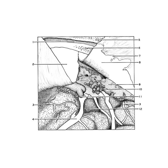
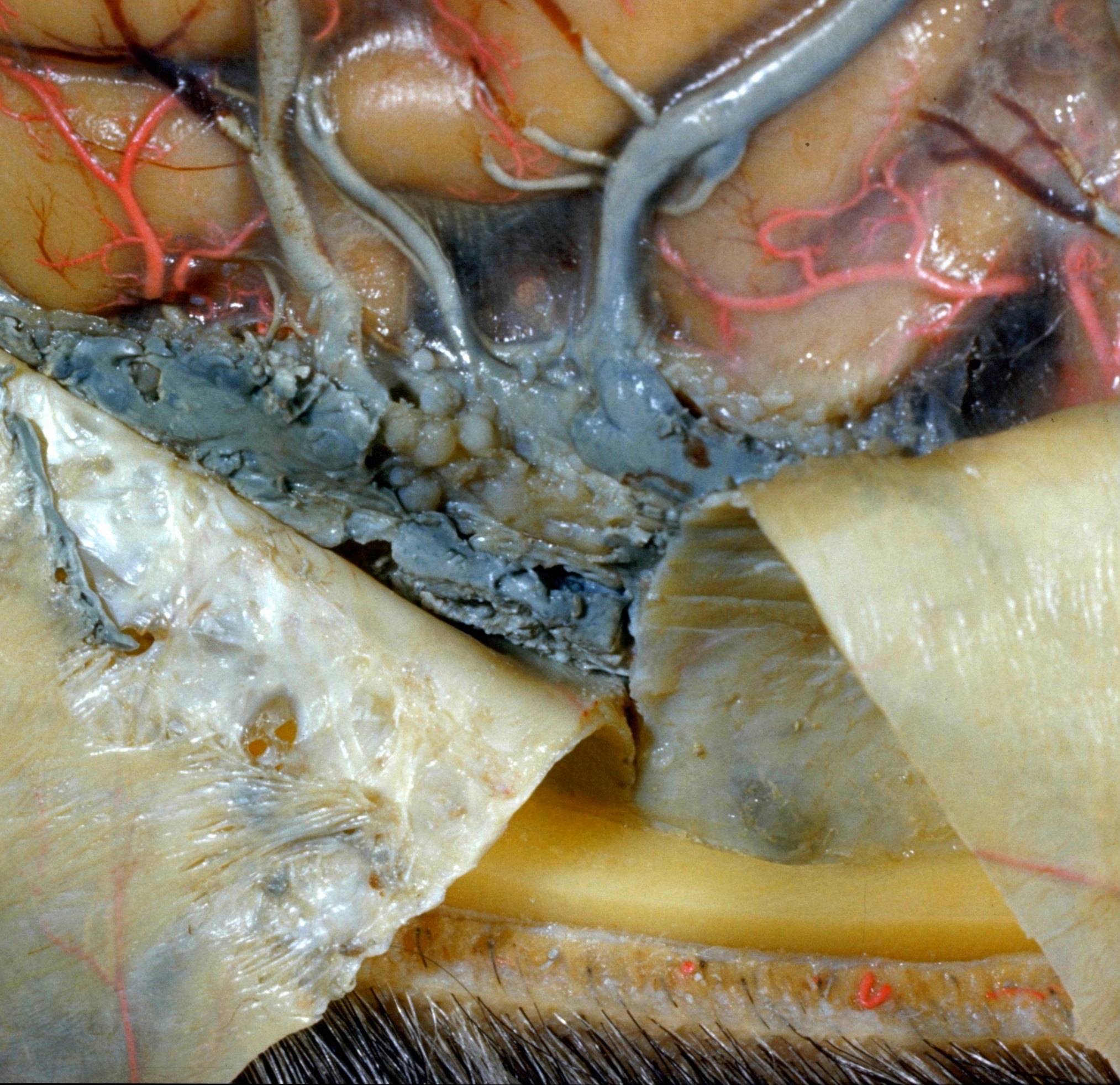
Exploration of the meninges and brain in situ
Dura reflected; arachnoid granulations projecting into venous lacunae
The dura has been incised and turned medially to open a large venous lacuna, the margin of which can be seen on the under surface of the dura at 7. The blue latex which fills the lacuna has been cut away enough to reveal a number of arachnoid granulations (10) which protrude into the venous pool. Note that several cortical veins appear to open directly into the venous lacuna. The cut surface of parietal bone (6) is in the midplane of the head. The superior sagittal sinus, which is not visible here, lies within the dura close to this bone. The arachnoid membrane remains over the cerebral hemisphere. The subarachnoid spaces within the sulci are visible. Several branches of the anterior cerebral artery are seen.
- External surface of parietal bone
- External sur face of dura mater
- Branch anterior cerebral artery
- Superior cerebral vein
- Superficial fascia
- Diploë in cut edge of parietal bone
- Lateral margin of venous lacuna (cut through and elevated)
- Endothelial surface of venus lacuna against dura mater
- Blue latex within lacuna
- Arachnoid granulations [Pacchioni]
- Point at which cerebral vein enters venous lacuna
- Arachnoid