X CLOSE

Volar aspect of forearm

Interosseous membrane

Stanford holds the copyright to the David L. Bassett anatomical images and has assigned Creative Commons license Attribution-Share Alike 4.0 International to all of the images.
For additional information regarding use and permissions, please contact Dr. Drew Bourn at dbourn@stanford.edu.

Image #99-7
Bassett ImageDiagram Image
Legend Image
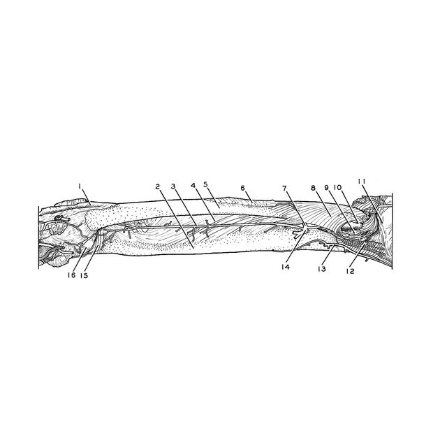
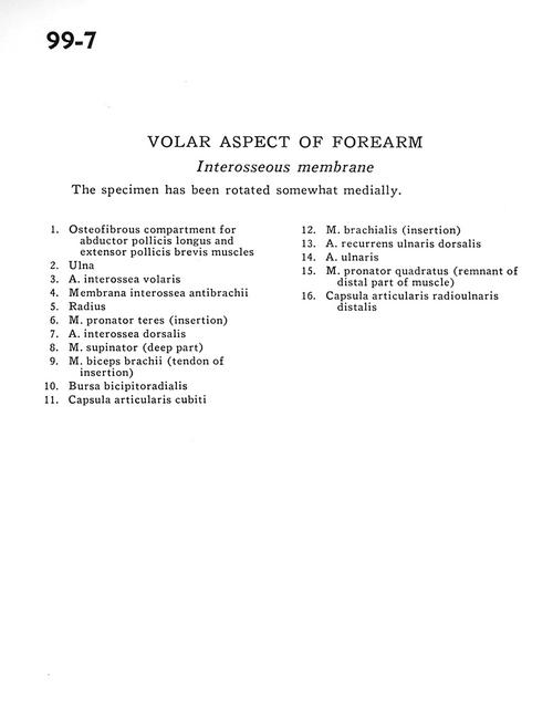
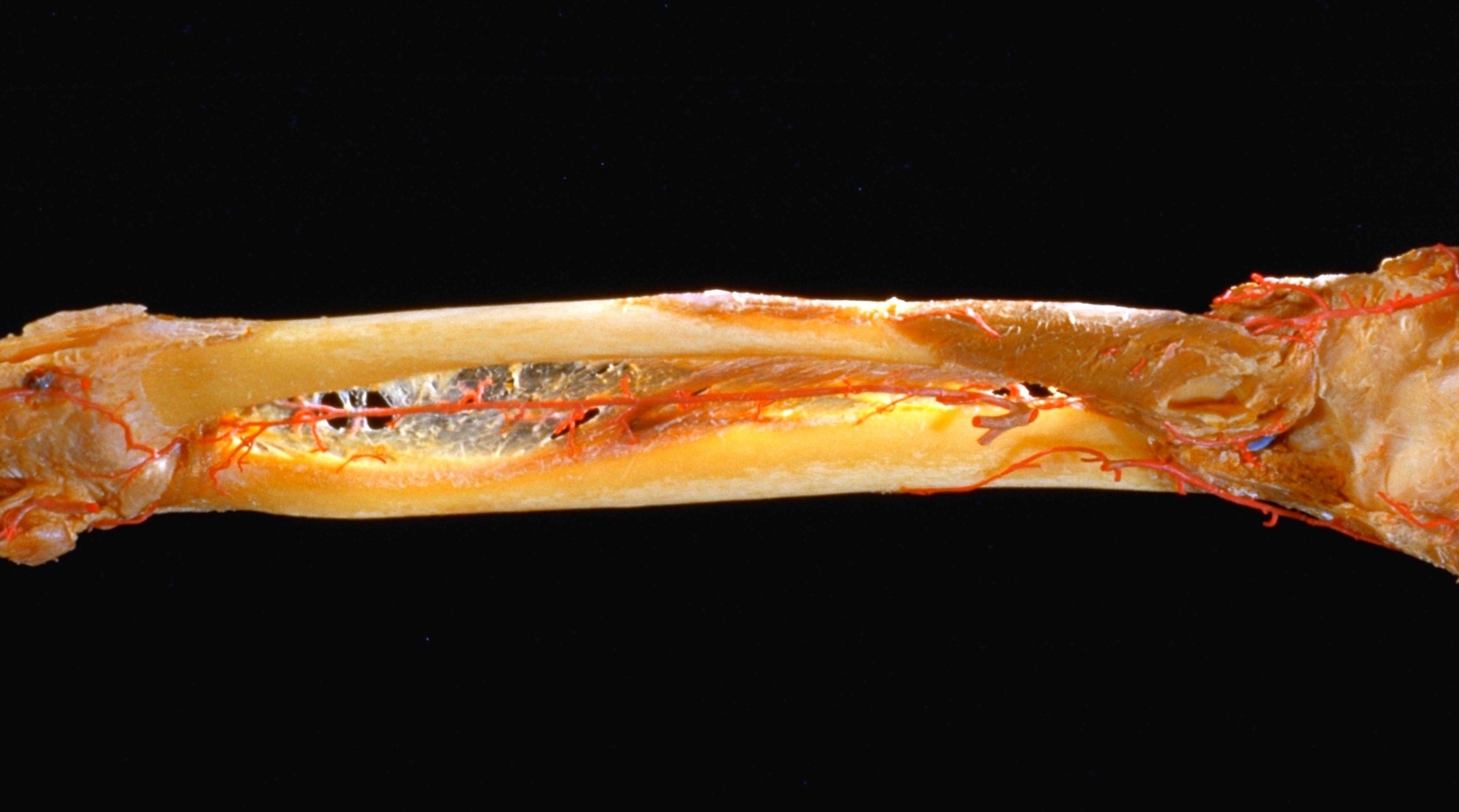
Volar aspect of forearm
Interosseous membrane
The specimen has been rotated somewhat medially.
1 . Osteofibrous compartment for abductor pollicis longus and extensor pollicis brevis muscles
2 . Ulna
3 . Anterior interosseous artery
4 . Antebrachial interosseous membrane
5 . Radius
6 . Pronator teres muscle (insertion)
7 . Dorsal interosseous artery
8 . Supinator muscle (deep part)
9 . Biceps brachii muscle (tendon of insertion)
10 . Bursa bicipitoradialis
11 . Cubital joint capsule
12 . Brachialis muscle (insertion)
13 . Dorsal recurrent ulnar artery
14 . Ulnar artery
15 . Pronator quadratus muscle (remnant of distal part of muscle)
16 . Distal radioulnar joint capsule