X CLOSE

Volar aspect of forearm

Nerve supply to flexor digitorum profundus muscle (continued)

Stanford holds the copyright to the David L. Bassett anatomical images and has assigned Creative Commons license Attribution-Share Alike 4.0 International to all of the images.
For additional information regarding use and permissions, please contact Dr. Drew Bourn at dbourn@stanford.edu.

Image #99-3
Bassett ImageDiagram Image
Legend Image
Volar aspect of forearm
Nerve supply to flexor digitorum profundus muscle (continued)
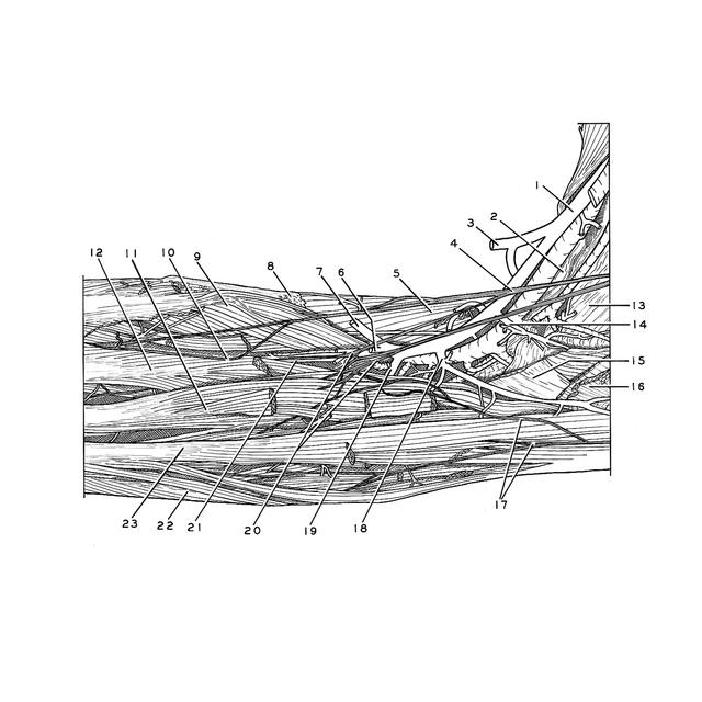
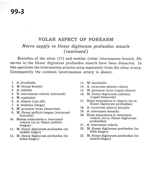
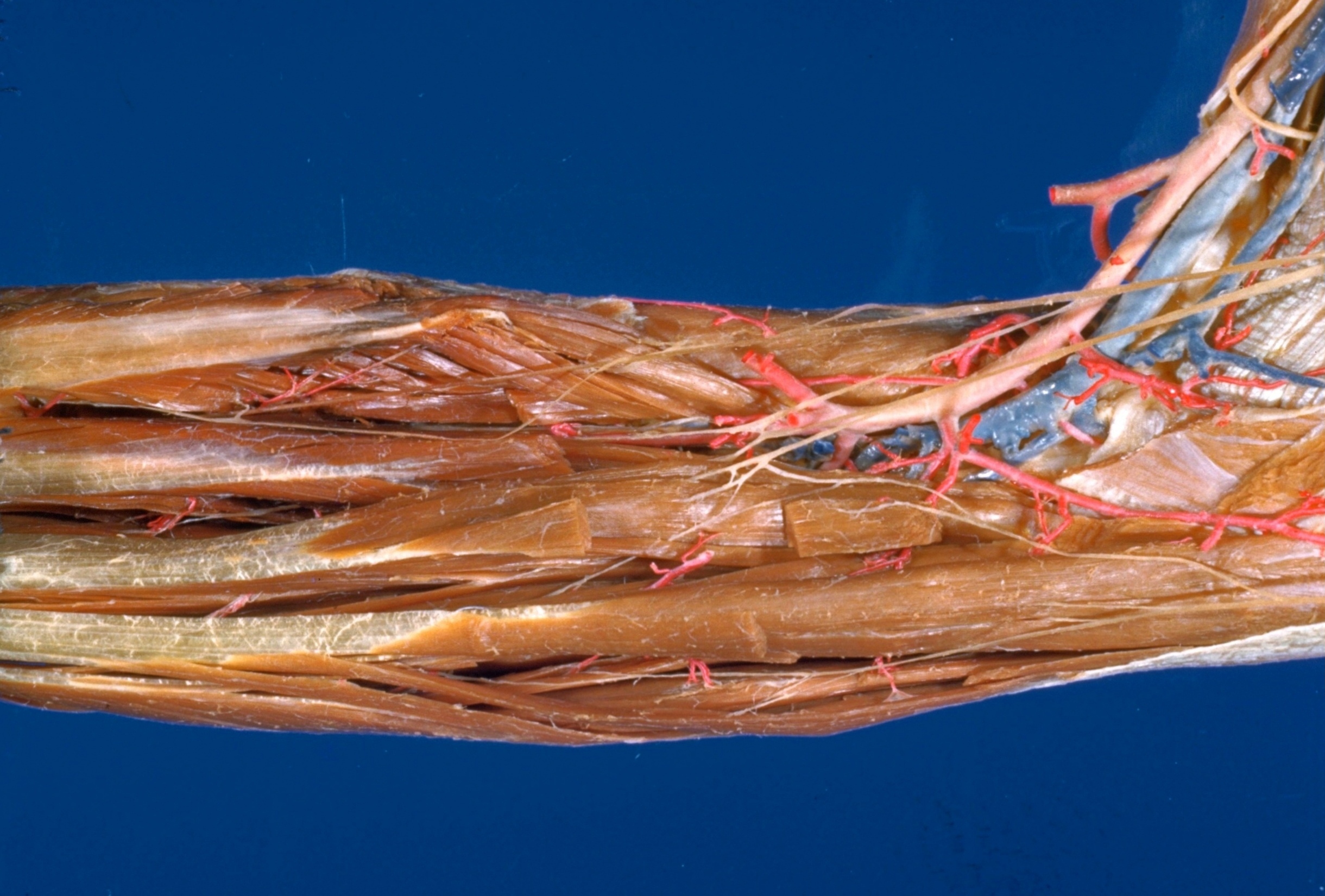
Branches of the ulnar (17) and median (volar interosseous branch, 20) nerves to the flexor digitorum profundus muscle have been dissected. In this specimen the interosseous arteries arise separately from the ulnar artery. Consequently the common interosseous artery is absent.
1 . Brachial artery
2 . Biceps brachii muscle
3 . Radial artery
4 . Anterior interosseous nerve (elevated)
5 . Supinator muscle
6 . Ulnar artery (cut off)
7 . Median artery (large)
8 . Pronator teres muscle (insertion)
9 . Flexor pollicis longus muscle (retracted laterally)
10 . Muscular branch anterior interosseous nerve (to flexor pollicis longus muscle)
11 . Flexor digitorum profundus muscle (to middle finger)
12 . Flexor digitorum profundus muscle (to index finger)
13 . Brachialis muscle
14 . Anterior recurrent ulnar artery
15 . Pronator teres muscle (ulnar head)
16 . Flexor digitorum superficialis (humeral head)
17 . Muscular branches of ulnar nerve (to flexor digitorum profundus muscle)
18 . Dorsal recurrent ulnar artery
19 . Dorsal interosseous artery
20 . Muscular branches anterior interosseous nerve (to flexor digitorum profundus muscle)
21 . Anterior interosseous artery
22 . Flexor digitorum profundus muscle (to fifth finger)
23 . Flexor digitorum profundus muscle fourth finger)