X CLOSE

Arm

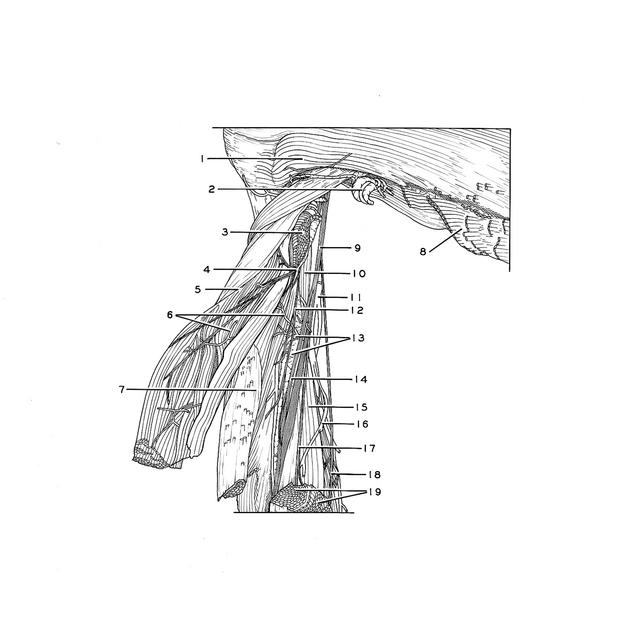
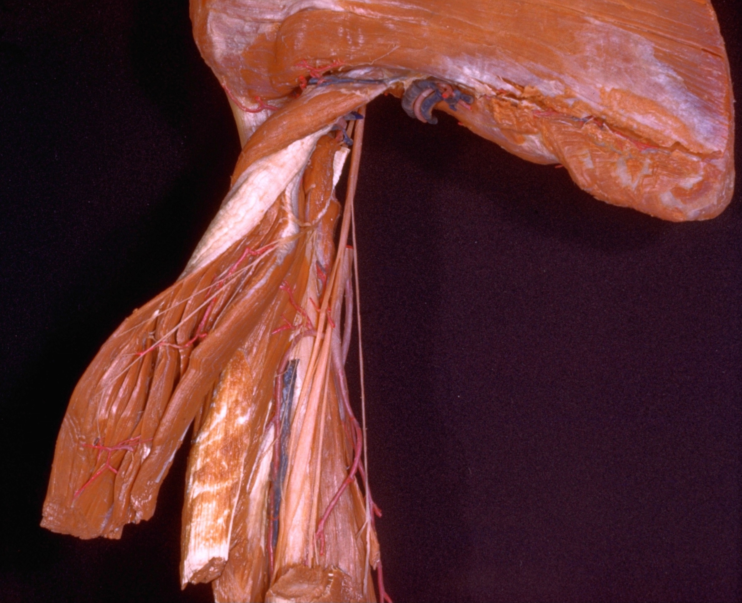
Relation of left radial nerve to triceps muscle, posterior view

Stanford holds the copyright to the David L. Bassett anatomical images and has assigned Creative Commons license Attribution-Share Alike 4.0 International to all of the images.
For additional information regarding use and permissions, please contact Dr. Drew Bourn at dbourn@stanford.edu.

Image #96-5
Bassett ImageDiagram Image
Legend Image
Arm
Relation of left radial nerve to triceps muscle, posterior view
The long and lateral heads of the triceps muscle have been transected (19) and reflected laterally.
1 . Teres minor muscle
2 . Circumflex scapular artery
3 . Teres major muscle
4 . Muscular branches of radial nerve
5 . Triceps brachii muscle (long head, reflected laterally)
6 . Muscular branches brachial artery
7 . Triceps brachii muscle (lateral head, reflected laterally; pointer on intramuscular aponeurosis which unites long and lateral heads of muscle)
8 . Teres major muscle (area of origin)
9 . Radial nerve (retracted medially)
10 . Coracobrachialis muscle (in background)
11 . Brachial artery
12 . Muscular branch of radial nerve (to lateral head of triceps)
13 . Upper pointer: Deep brachial artery (continues distally as radial collateral artery) Lower pointer: Body of humerus (covered by periosteum pointer on sulcus of radial nerve)
14 . Dorsal antebrachial cutaneous nerve
15 . Triceps brachii muscle (medial head)
16 . Superior ulnar collateral artery
17 . Muscular branch of radial nerve (to lower part of medial head of triceps and to anconeus muscle)
18 . Muscular branch of radial nerve (to medial head of triceps)
19 . Triceps brachii muscle (transected; upper pointer, lateral head; lower pointer, long head)