X CLOSE

Shoulder

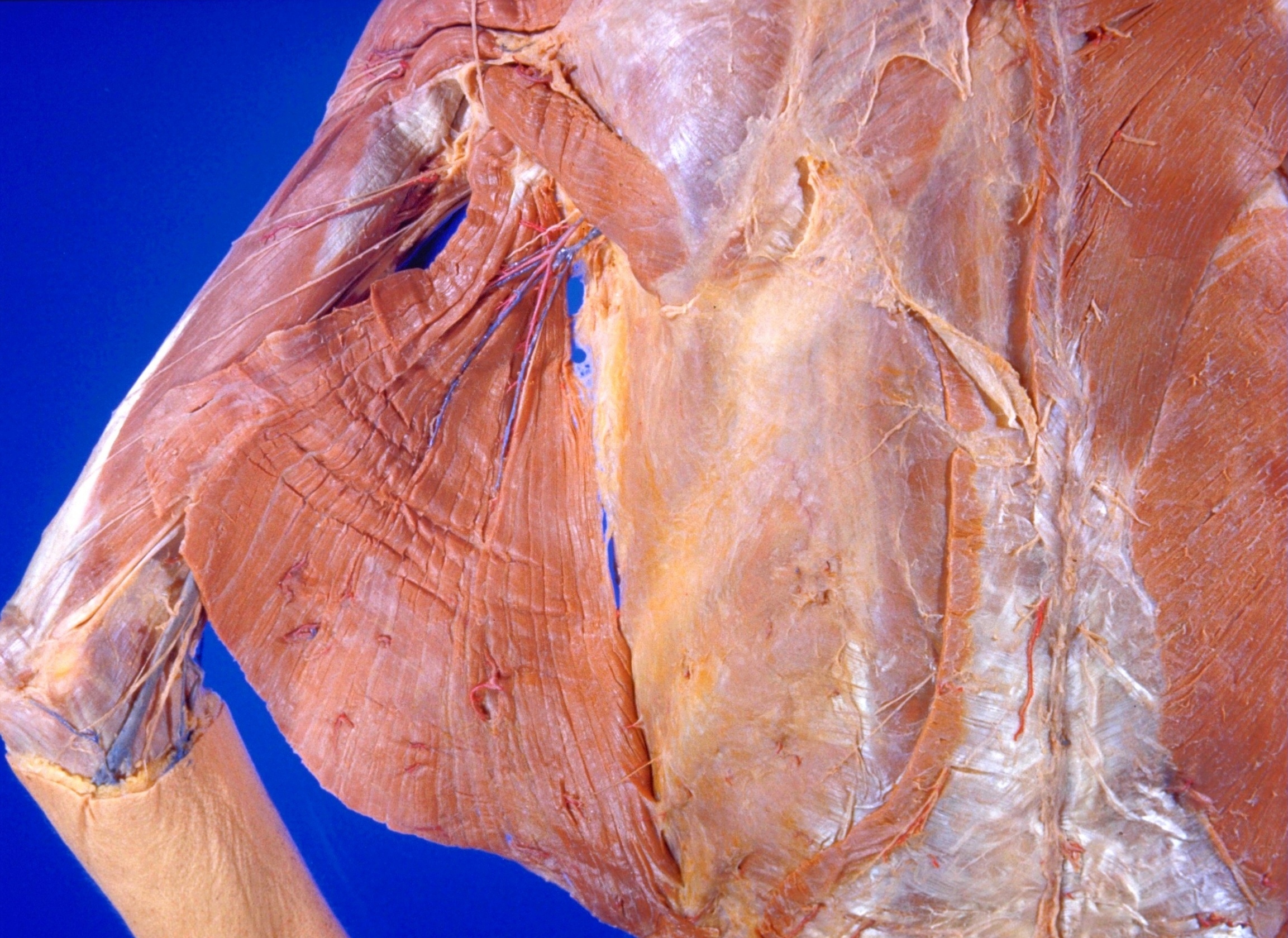
Nerve and blood supply to left lattisimus dorsi muscle

Stanford holds the copyright to the David L. Bassett anatomical images and has assigned Creative Commons license Attribution-Share Alike 4.0 International to all of the images.
For additional information regarding use and permissions, please contact Dr. Drew Bourn at dbourn@stanford.edu.

Image #93-3
Bassett ImageDiagram Image
Legend Image
Shoulder
Nerve and blood supply to left lattisimus dorsi muscle
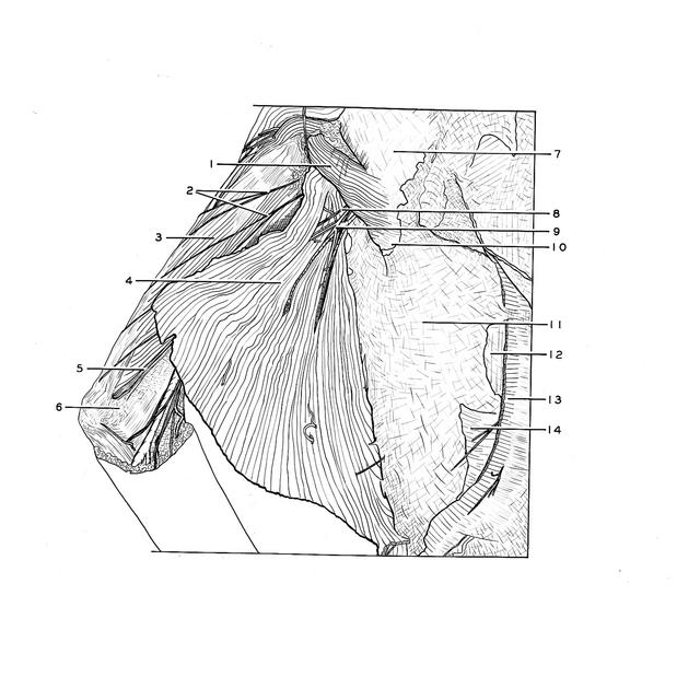
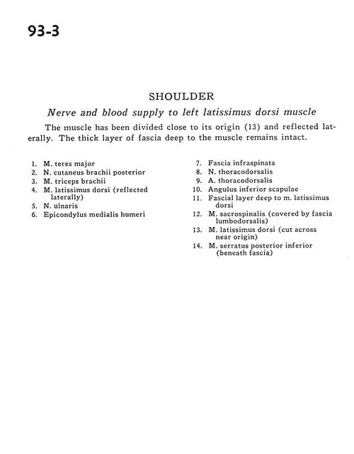
The muscle has been divided close to its origin (13) and reflected laterally. The thick layer of fascia deep to the muscle remains intact.
1 . Teres major muscle
2 . Posterior brachial cutaneous nerve
3 . Triceps brachii muscle
4 . Latissimus dorsi muscle (reflected laterally)
5 . Ulnar nerve
6 . Medial epicondyle of humerus
7 . Infraspinatus fascia
8 . Thoracodorsal nerve
9 . Thoracodorsal artery
10 . Inferior angle of scapula
11 . Fascial layer deep to latissimus dorsi muscle
12 . Sacrospinalis muscle (covered by lumbodorsal fascia)
13 . Latissimus dorsi muscle (cut across near origin)
14 . Posterior inferior serratus muscle (beneath fascia)