X CLOSE

Dissection of head and neck from a posterior approach

Cavity of larynx, superior view

Stanford holds the copyright to the David L. Bassett anatomical images and has assigned Creative Commons license Attribution-Share Alike 4.0 International to all of the images.
For additional information regarding use and permissions, please contact Dr. Drew Bourn at dbourn@stanford.edu.

Image #83-4
Bassett ImageDiagram Image
Legend Image
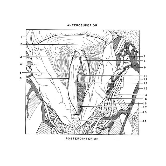
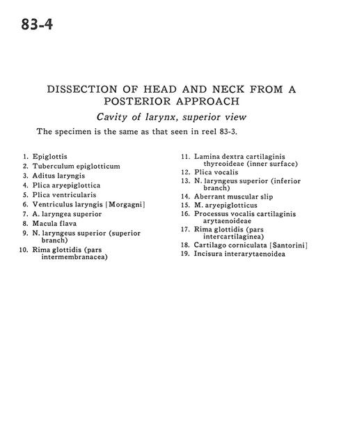
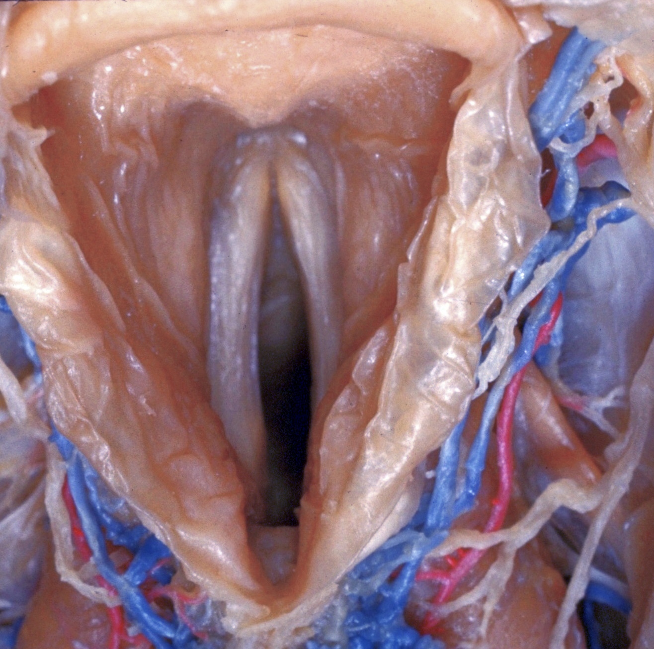
Dissection of head and neck from a posterior approach
Cavity of larynx, superior view
The specimen is the same as that seen in reel 83-3.
1 . Epiglottis
2 . Epiglottic tubercle
3 . Laryngeal ventricle
4 . Aryepiglottic fold
5 . Ventricular fold
6 . Laryngeal ventricle
7 . Superior laryngeal artery
8 . Macula flava
9 . Superior laryngeal nerve (superior branch)
10 . Glottic rim (intermembrane part )
11 . Lamina right thyroid cartilage (inner surface)
12 . Vocal cord
13 . Superior laryngeal nerve (inferior branch)
14 . Aberrant muscular slip
15 . Aryepiglotticus muscle
16 . Vocal process of arytenoid cartilage
17 . Glottic rim (intercartilaginous part)
18 . Corniculate cartilage
19 . Interarytenoid notch