Dissection of head and neck from a posterior approach
General view of pharynx and esophagus
For permissions information regarding the use of these images,
please contact Drew Bourn at dbourn@stanford.edu
Image #82-2
Dissection of head and neck from a posterior approach
General view of pharynx and esophagus
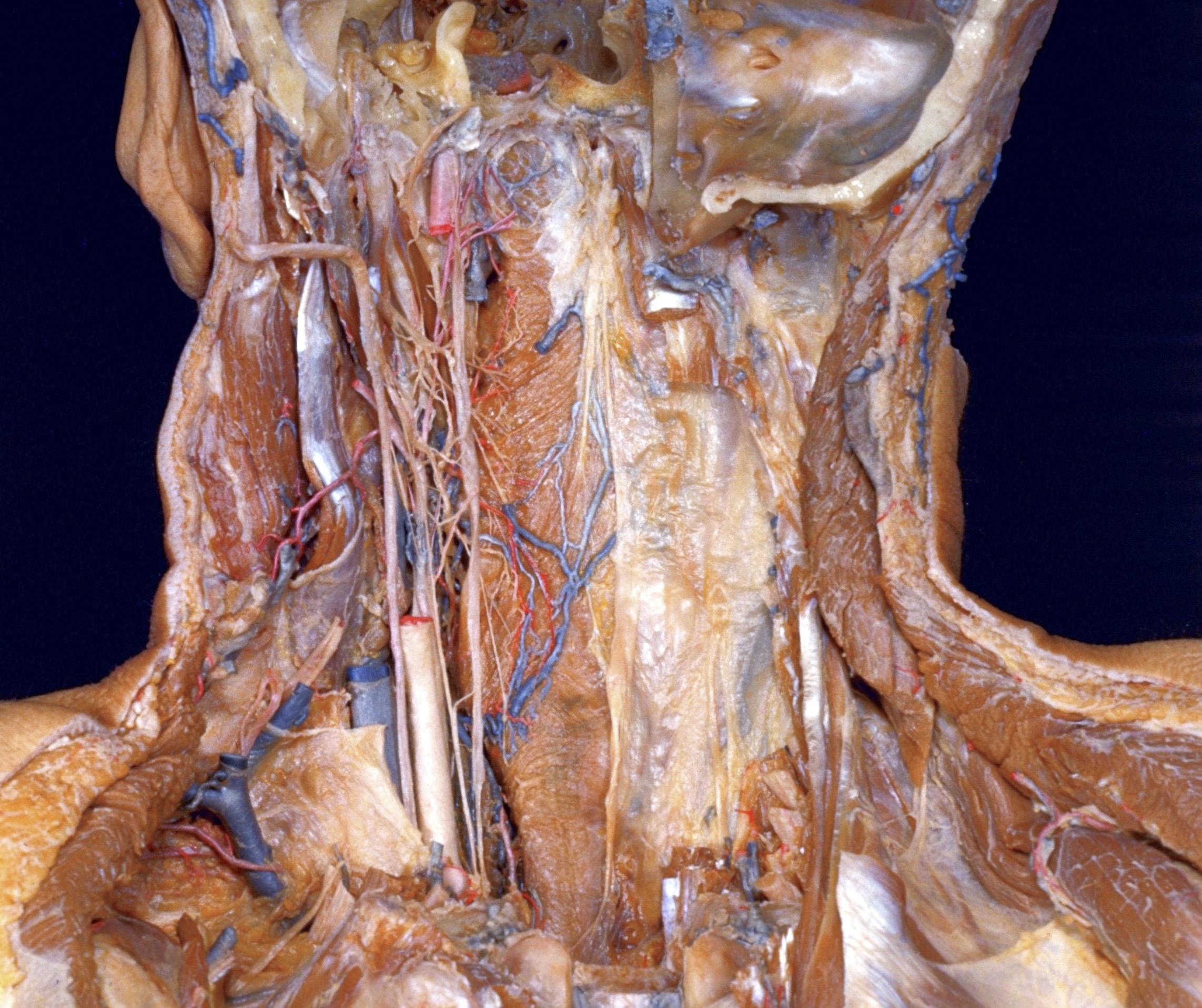
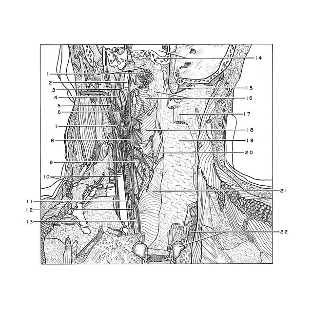
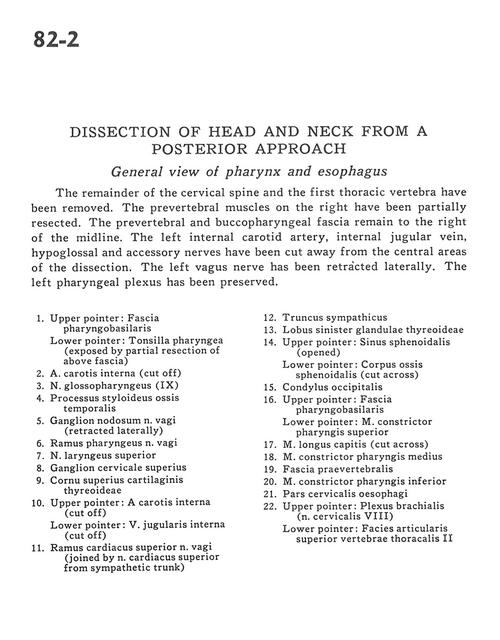
The remainder of the cervical spine and the first thoracic vertebrae have been removed. The prevertebral muscles on the right have been partially resected. The prevertebral and buccopharyngeal fascia remain to the right of the midline. The left internal carotid artery, internal jugular vein, hypoglossal and accessory nerves have been cut away from the central areas of the dissection. The left vagus nerve has been retracted laterally. The left pharyngeal plexus has been preserved.
1
.
Upper pointer: Pharyngobasilar fascia Lower pointer: Pharyngeal tonsil (exposed by partial resection of above fascia)
2
.
Internal carotid artery (cut off)
3
.
Glossopharyngeal nerve (IX)
4
.
Styloid process temporal bone
5
.
Nodose ganglion of vagus nerve (retracted laterally)