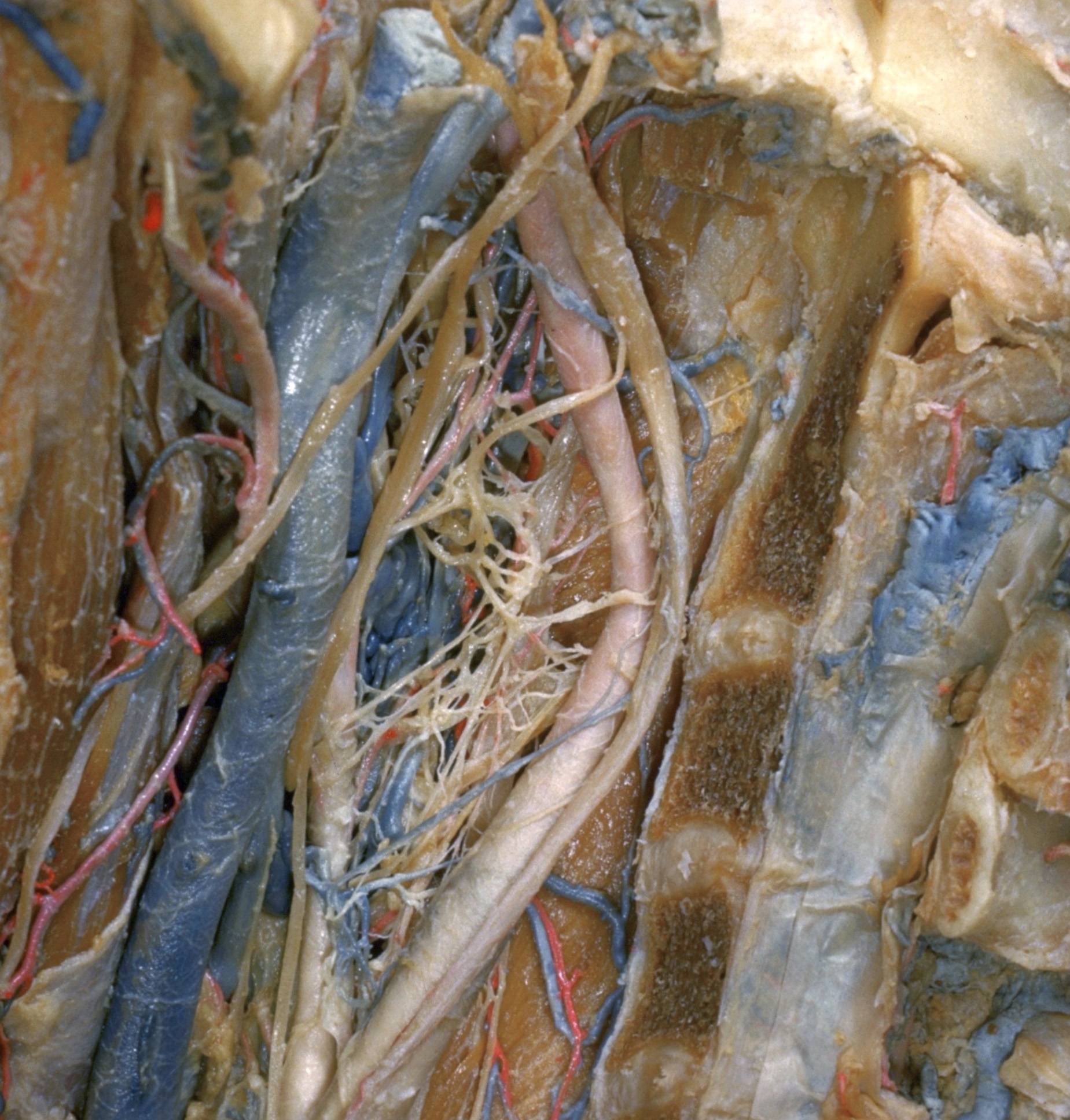
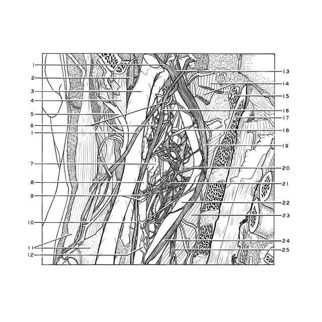
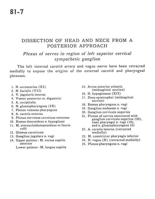
Dissection of head and neck from a posterior approach
Plexus of nerves in region of left superior cervical sympathetic ganglion
Stanford holds the copyright to the David L. Bassett anatomical images and has assigned
Creative Commons license Attribution-Share Alike 4.0 International to all of the images.
For additional information regarding use and permissions,
please contact Dr. Drew Bourn at dbourn@stanford.edu.
Image #81-7
 |  | ||||||||||||||||||||||||||||||||||||||||||||||||||||||
 |
|