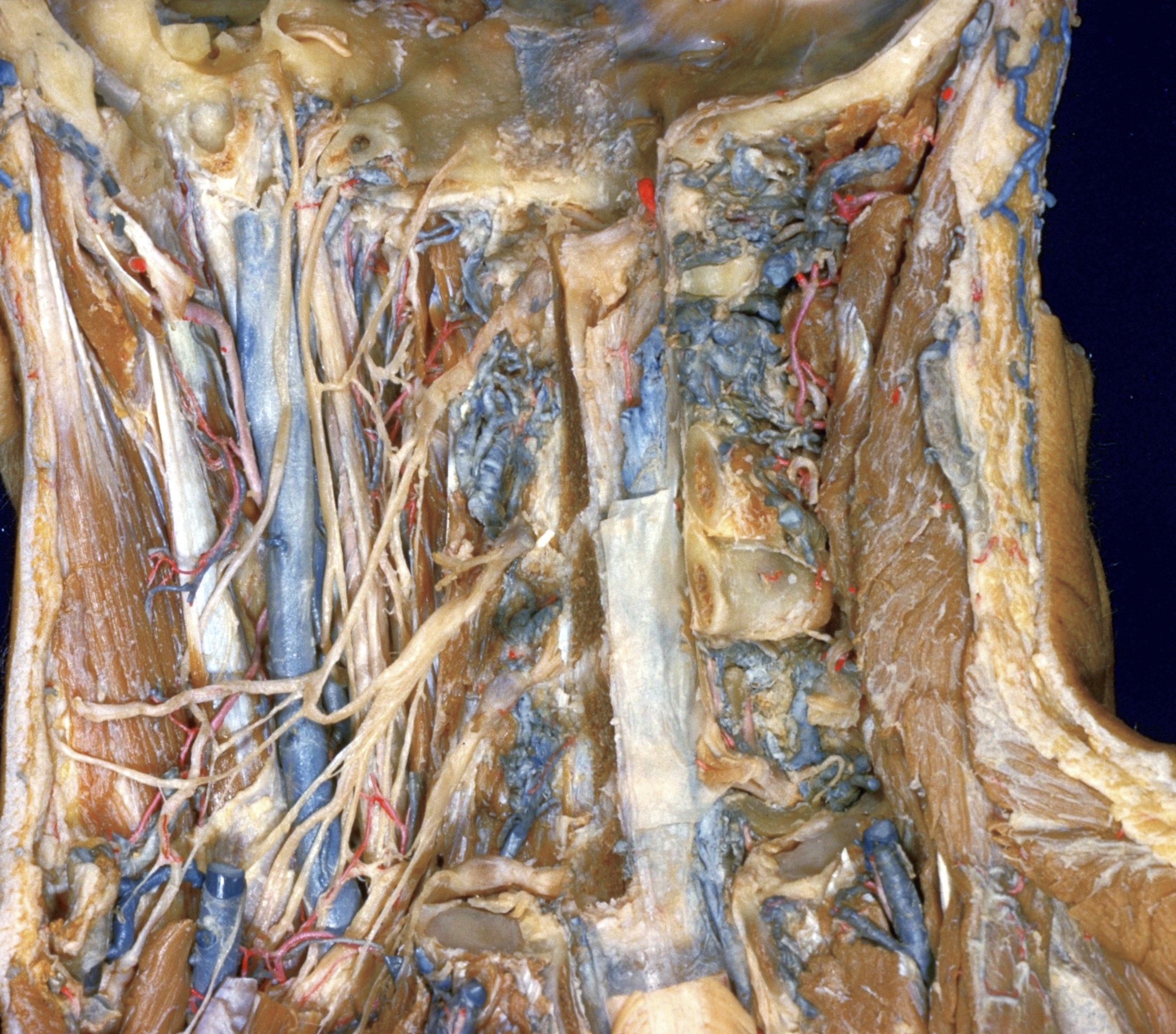
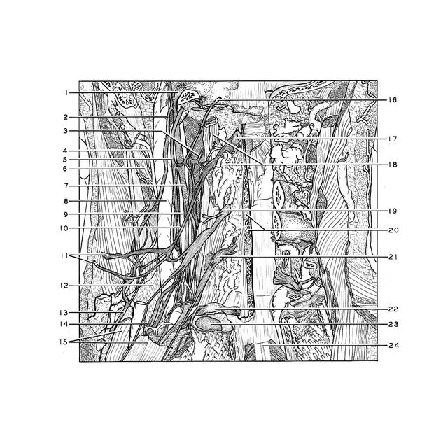
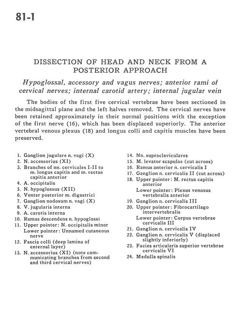
Dissection of head and neck from a posterior approach
Hypoglossal, accessory and vagus nerves; anterior rami of cervical nerves; internal carotid artery; internal jugular vein
Stanford holds the copyright to the David L. Bassett anatomical images and has assigned
Creative Commons license Attribution-Share Alike 4.0 International to all of the images.
For additional information regarding use and permissions,
please contact Dr. Drew Bourn at dbourn@stanford.edu.
Image #81-1
 |  | ||||||||||||||||||||||||||||||||||||||||||||||||||||
 |
|