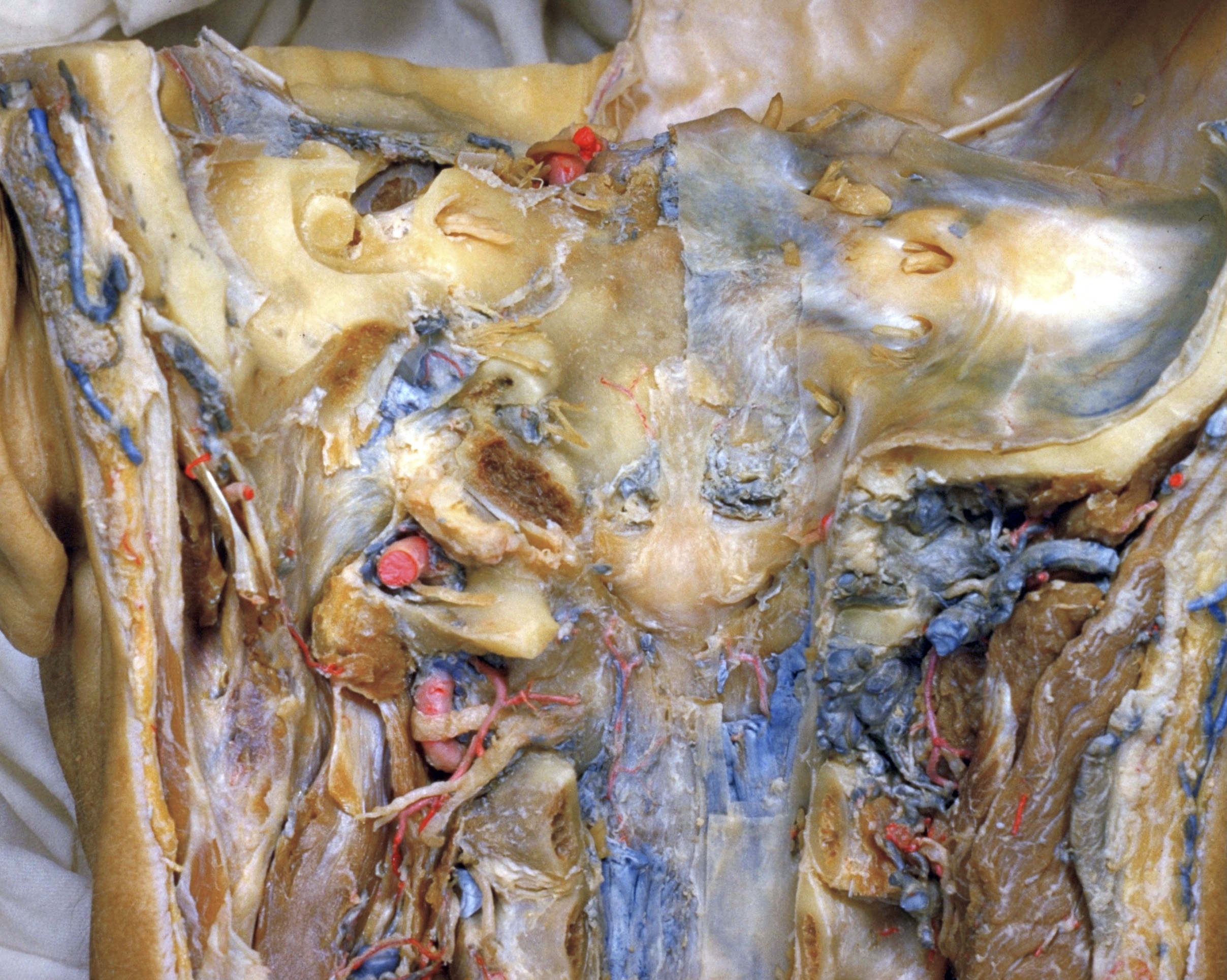
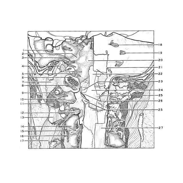
Dissection of head and neck from a posterior approach
Cruciate ligament; alar ligaments
For permissions information regarding the use of these images,
please contact Drew Bourn at dbourn@stanford.edu
Image #80-4
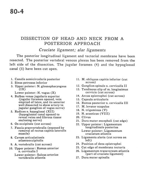
Dissection of head and neck from a posterior approach
Cruciate ligament; alar ligaments
The posterior longitudinal ligament and tectorial membrane have been resected. The posterior vertebral venous plexus has been removed from the left side of the dissection. The jugular foramen (4) and the hypoglossal canal (5) have been cut open.
Superior jugular venous bulb (jugular foramen opened, vein emptied of latex, and its anterior wall dissected to show artery to jugular ganglion of vagus nerve)
5
.
Roots of hypoglossal nerve (XII) (hypoglossal canal opened to reveal veins and fibrous tissue enclosing nerve)
6
.
Occipital bone (cut across)
7
.
Prevertebral fascia (exposed by removal of rectus capitis lateralis muscle)
8
.
Atlantooccipital cavity of articulation
9
.
Vertebral artery (cut across)
10
.
Upper pointer: Anterior branch of cervical nerve I Lower pointer: Groove in atlas for vertebral artery
11
.
Inferior oblique capitis muscle (cut across)
12
.
Spinal ganglion cervical nerve II
13
.
Tendinous slip of longissimus cervicis muscle (cut across)