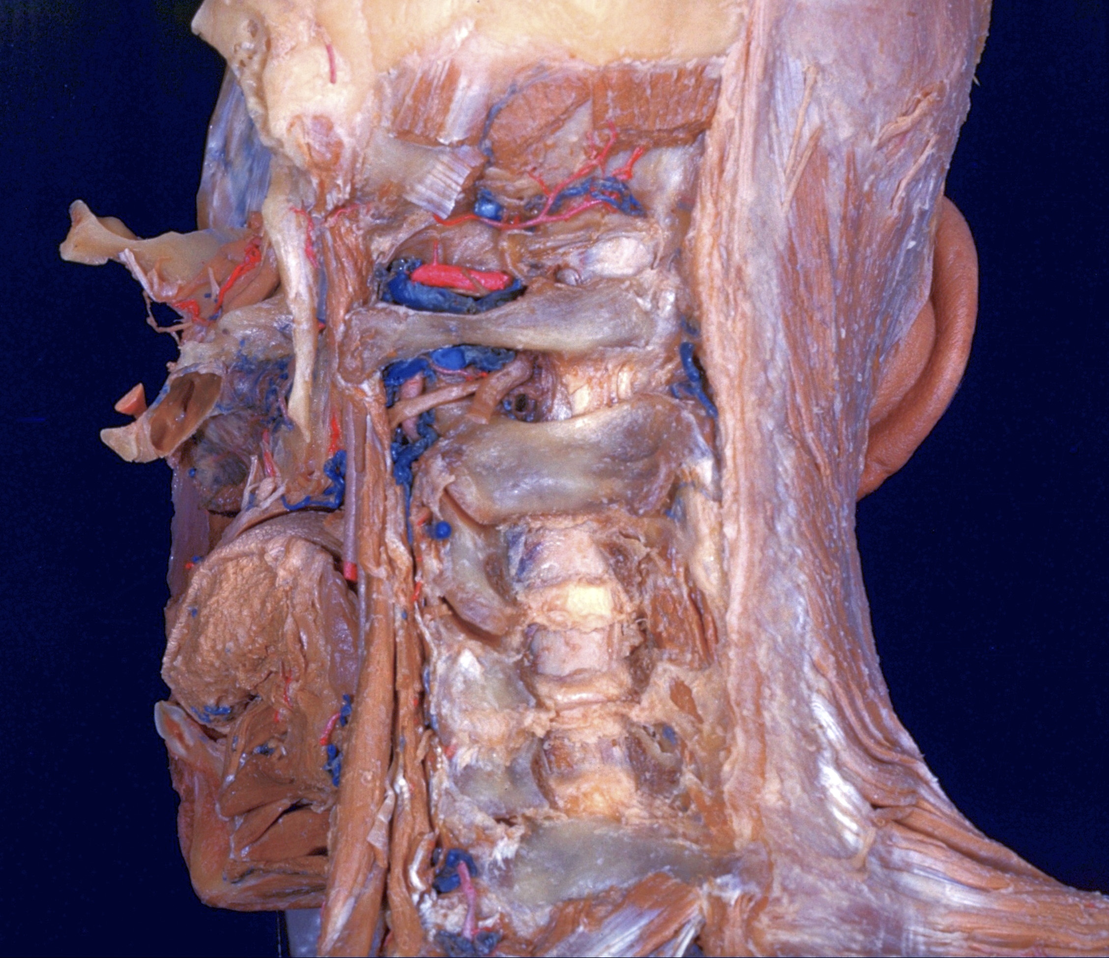
Dissection of head and neck from a posterior approach
Ligamenta flava
For permissions information regarding the use of these images,
please contact Drew Bourn at dbourn@stanford.edu
Image #80-1
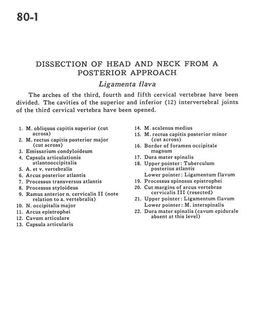
Dissection of head and neck from a posterior approach
Ligamenta flava
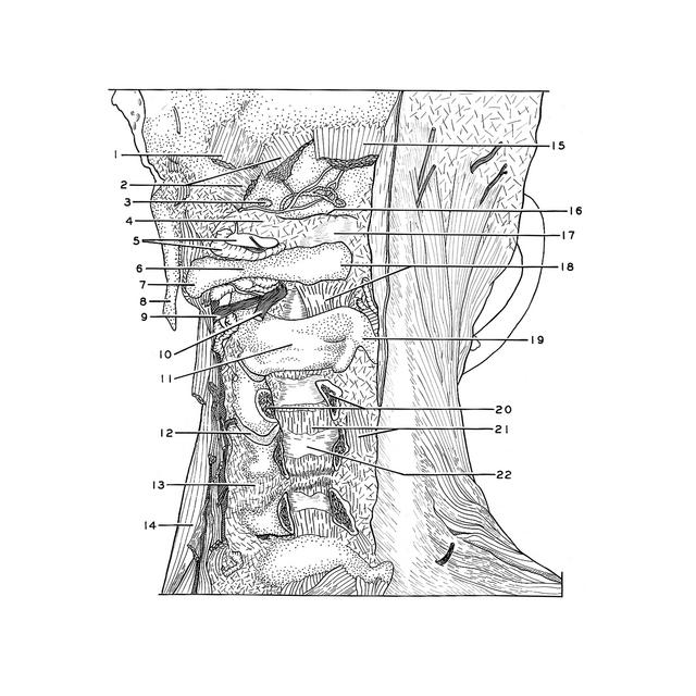
The arches of the third, fourth and fifth cervical vertebrae have been divided. The cavities of the superior and inferior (12) intervertebral joints of the third cervical vertebrae have been opened.
1
.
Superior oblique capitis muscle (cut across)
2
.
Major posterior rectus capitis muscle (cut across)
3
.
Condyloid emissary
4
.
Atlanto-occipital joint capsule
5
.
Vertebral artery and vein
6
.
Posterior arch of atlas
7
.
Transverse process atlas
8
.
Styloid process
9
.
Anterior branch of cervical nerve II (note relation to vertebral artery)
10
.
Greater occipital nerve
11
.
Arch axis
12
.
Articular cavity
13
.
Joint capsule
14
.
Middle scalene muscle
15
.
Minor posterior rectus capitis muscle (cut across)