For permissions information regarding the use of these images,
please contact Drew Bourn at dbourn@stanford.edu
Image #8-3
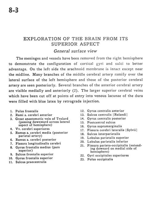
Exploration of the brain from its superior aspect
General surface view
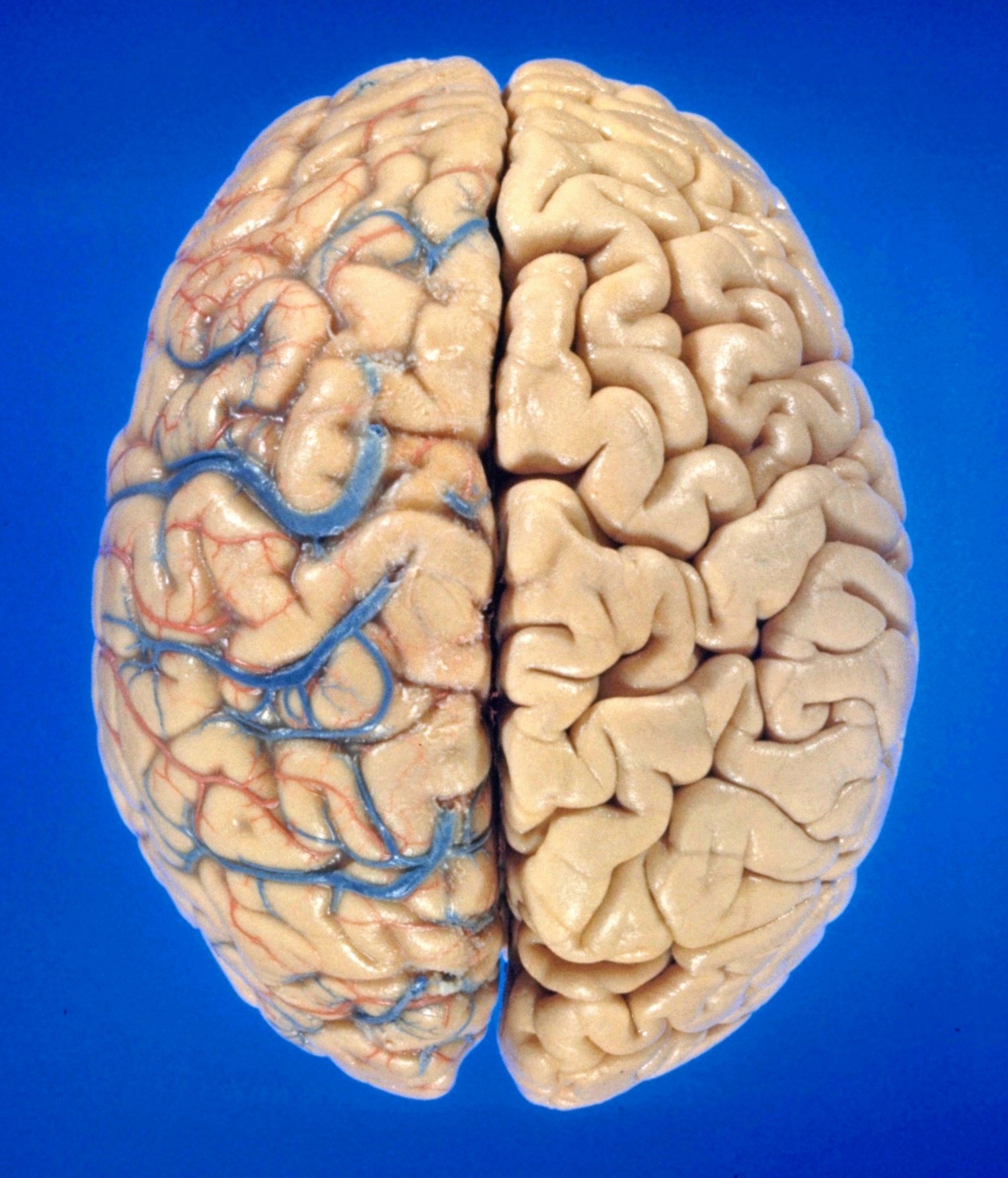
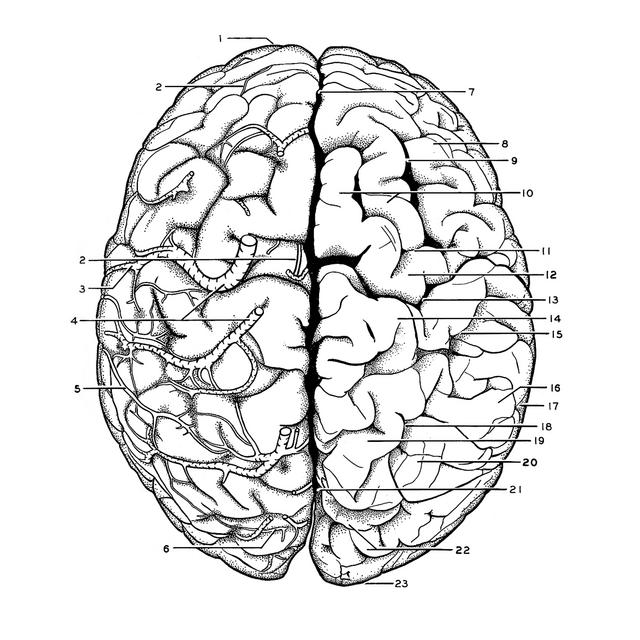
The meninges and vessels have been removed from the right hemisphere to demonstrate the configuration of cortical gyri and sulci to better advantage. On the left side the arachnoid membrane is intact except near the midline. Many branches of the middle cerebral artery ramify over the lateral surface of the left hemisphere and those of the posterior cerebral artery are seen posteriorly. Several branches of the anterior cerebral artery are visible medially and anteriorly (2). The larger superior cerebral veins which have been cut off at points of entry into venous lacunae of the dura were filled with blue latex by retrograde injection.
1
.
Frontal pole
2
.
Branch anterior cerebral artery
3
.
Great anastomotic vein of Trolard (passing downward across lateral aspect of hemisphere)