X CLOSE

Dissection of anterior and lateral regions of neck

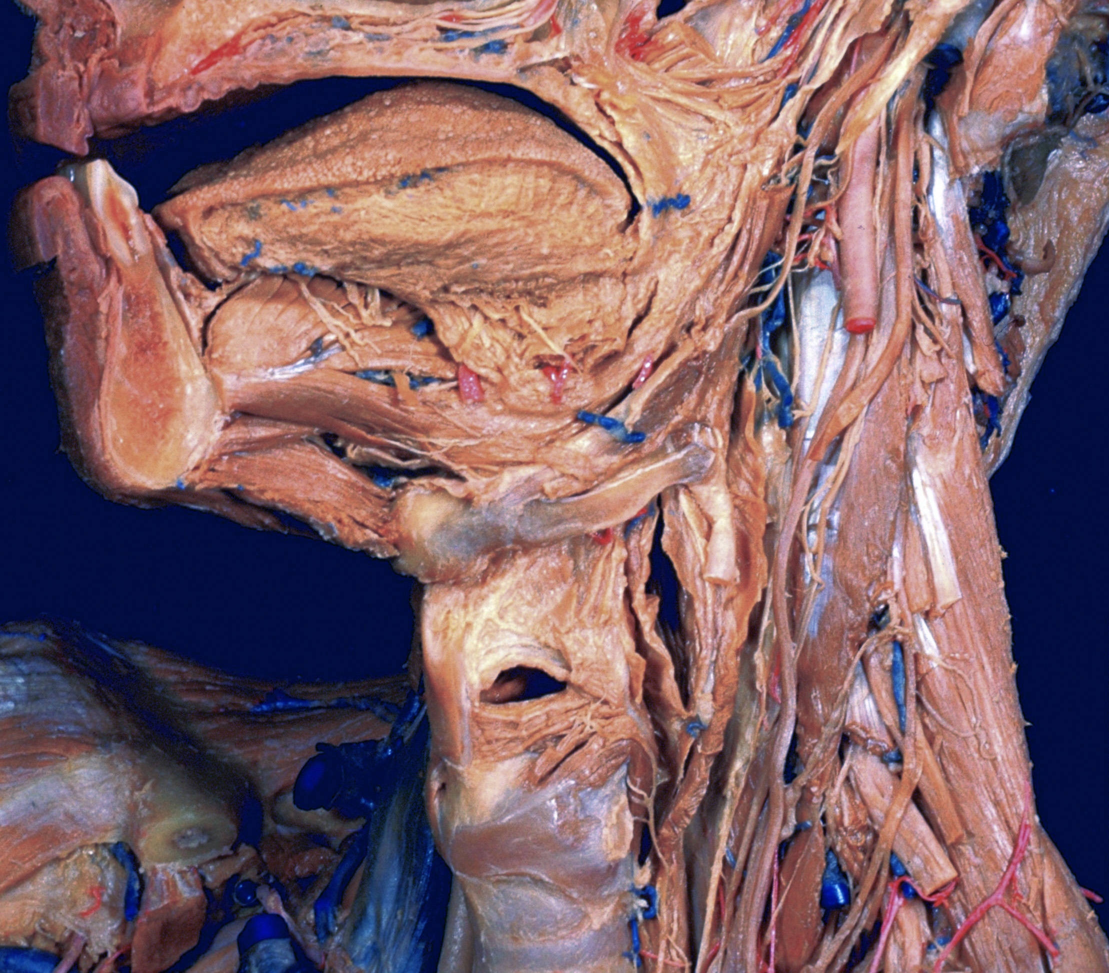
Larynx; relation to tongue, pharynx and hyoid bone, left anterolateral view

Stanford holds the copyright to the David L. Bassett anatomical images and has assigned Creative Commons license Attribution-Share Alike 4.0 International to all of the images.
For additional information regarding use and permissions, please contact Dr. Drew Bourn at dbourn@stanford.edu.

Image #77-6
Bassett ImageDiagram Image
Legend Image
Dissection of anterior and lateral regions of neck
Larynx; relation to tongue, pharynx and hyoid bone, left anterolateral view
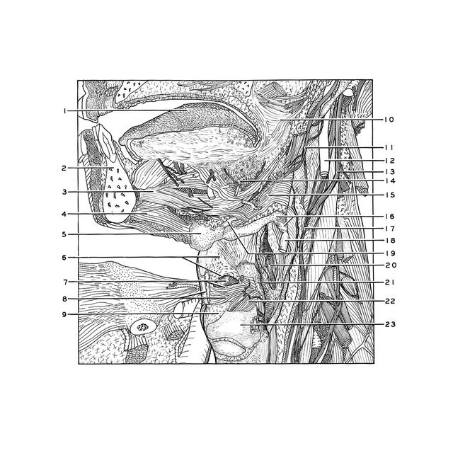
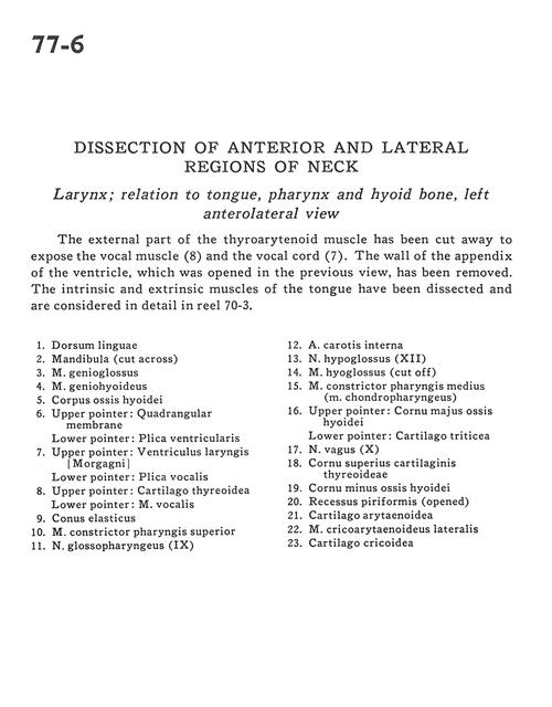
The external part of the thyroarytenoid muscle has been cut away to expose the vocal muscle (8) and the vocal cord (7). The wall of the appendix of the ventricle, which was opened in the previous view, has been removed. The intrinsic and extrinsic muscles of the tongue have been dissected and are considered in detail in reel 70-3.
1 . Dorsum of tongue
2 . Mandible (cut across)
3 . Genioglossus muscle
4 . Geniohyoid muscle
5 . Body hyoid bone
6 . Upper pointer: Quadrangular membrane Lower pointer: Ventricular fold
7 . Upper pointer: Laryngeal ventricle Lower pointer: Vocal cord
8 . Upper pointer: Thyroid cartilage Lower pointer: Vocalis muscle
9 . Conus elasticus
10 . Superior pharyngeal constrictor muscle
11 . Glossopharyngeal nerve (IX)
12 . Internal carotid artery
13 . Hypoglossal nerve (XII)
14 . Hyoglossus muscle (cut off)
15 . Middle pharyngeal constrictor muscle (chondropharyngeus)
16 . Upper pointer: Greater horn hyoid bone Lower pointer: Triticeal cartilage
17 . Vagus nerve (X)
18 . Superior horn thyroid cartilage
19 . Lesser horn hyoid bone
20 . Piriform recess (opened)
21 . Arytenoid cartilage
22 . Lateral cricoarytenoid muscle
23 . Cricoid cartilage