X CLOSE

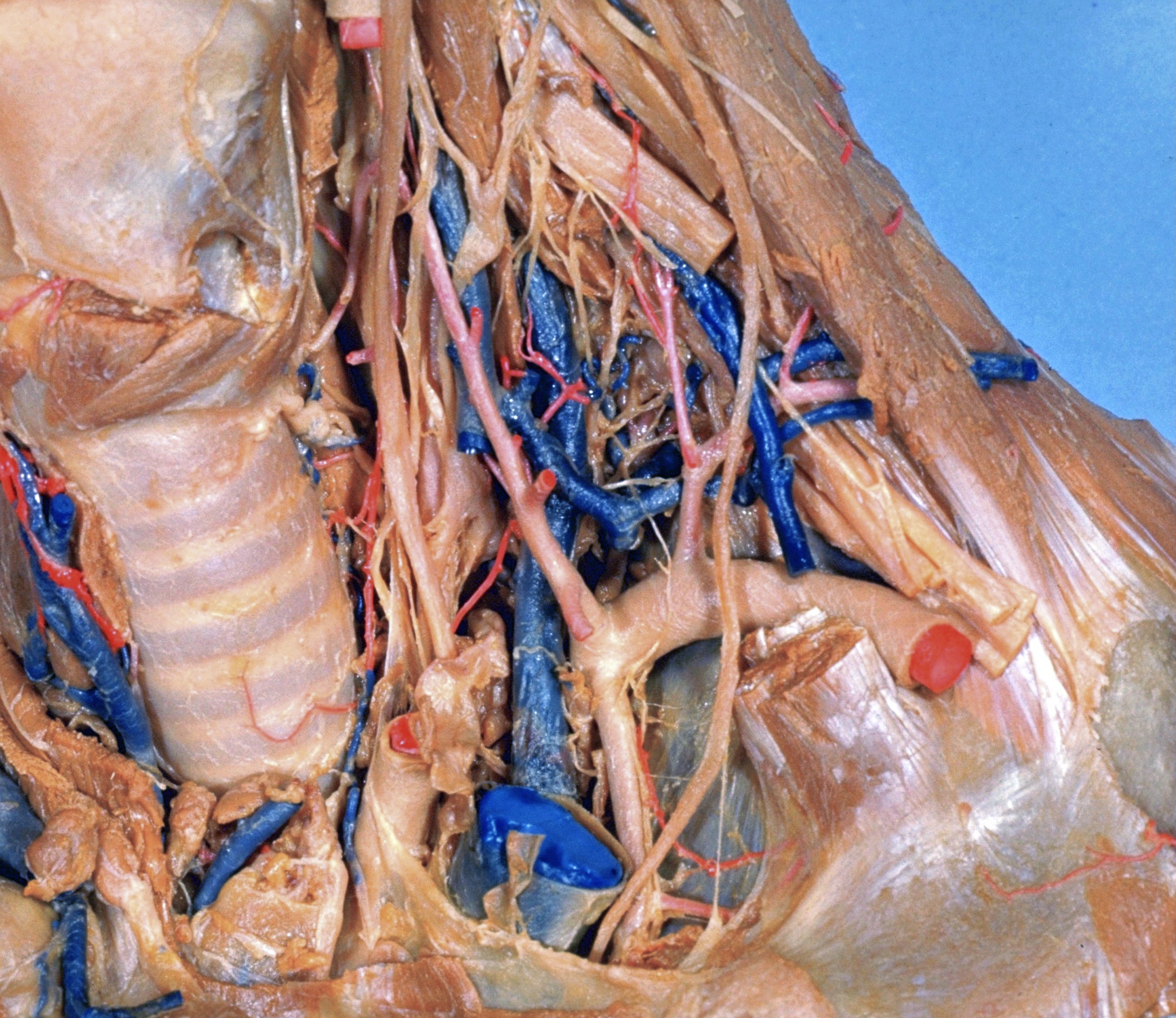
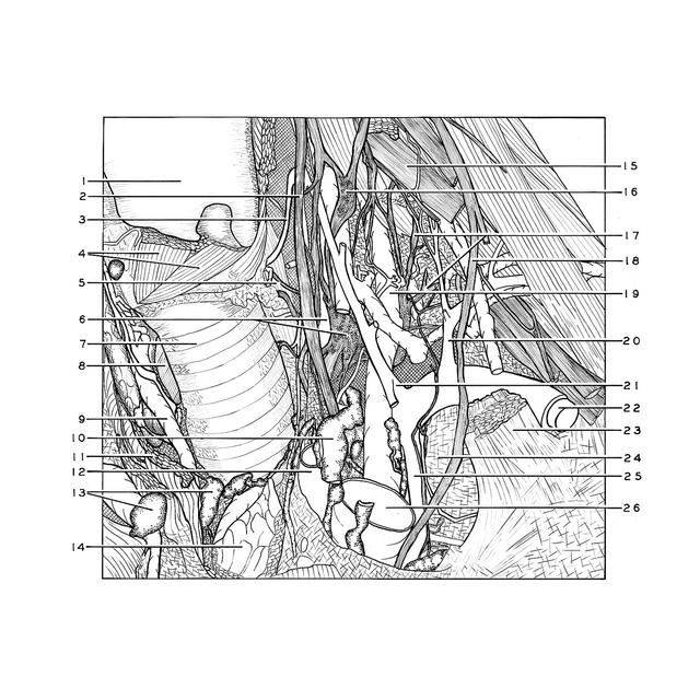
Dissection of anterior and lateral regions of neck

Relations of cupula of pleura, left anterolateral view

For permissions information regarding the use of these images, please contact Drew Bourn at dbourn@stanford.edu

Image #76-5
Bassett ImageDiagram Image
Legend Image
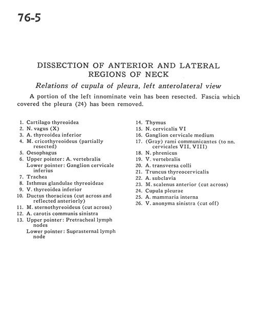
Dissection of anterior and lateral regions of neck
Relations of cupula of pleura, left anterolateral view
A portion of the left innominate vein has been resected. Fascia which covered the pleura (24) has been removed.
1 . Thyroid cartilage
2 . Vagus nerve (X)
3 . Inferior thyroid artery
4 . Cricothyroid muscle(partially resected)
5 . Esophagus
6 . Upper pointer: Vertebral artery Lower pointer: Inferior cervical ganglion
7 . Trachea
8 . Isthmus of thyroid gland
9 . Inferior thyroid vein
10 . Thoracic duct (cut across and reflected anteriorly)
11 . Sternothyroid muscle (cut across)
12 . Common carotid artery left
13 . Upper pointer: Pretracheal lymph node Lower pointer: Suprasternal lymph node
14 . Thymus
15 . Cervical nerve VI
16 . Middle cervical ganglion
17 . (Gray) rami communicantes (to cervical nerve VII, VIII)
18 . Phrenic nerve
19 . Vertebral vein
20 . Transverse superficial artery
21 . Thyrocervical trunk
22 . Subclavian artery
23 . Anterior scalene muscle (cut across)
24 . Cupula pleurae
25 . Internal thoracic (mammary) artery
26 . Brachiocephalic vein left (cut off)