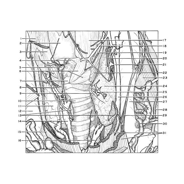
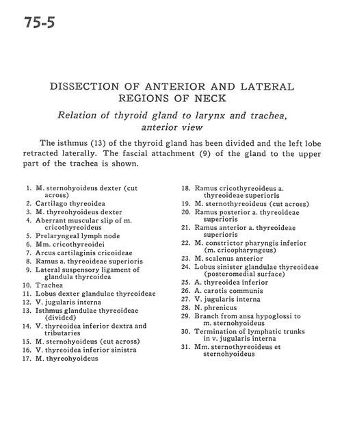
Dissection of anterior and lateral regions of neck
Relations of thyroid gland to larynx and trachea, anterior view
Stanford holds the copyright to the David L. Bassett anatomical images and has assigned
Creative Commons license Attribution-Share
Alike 4.0 International to all of the images.
For additional information regarding use and permissions,
please contact Dr. Drew Bourn at dbourn@stanford.edu.
Image #75-5
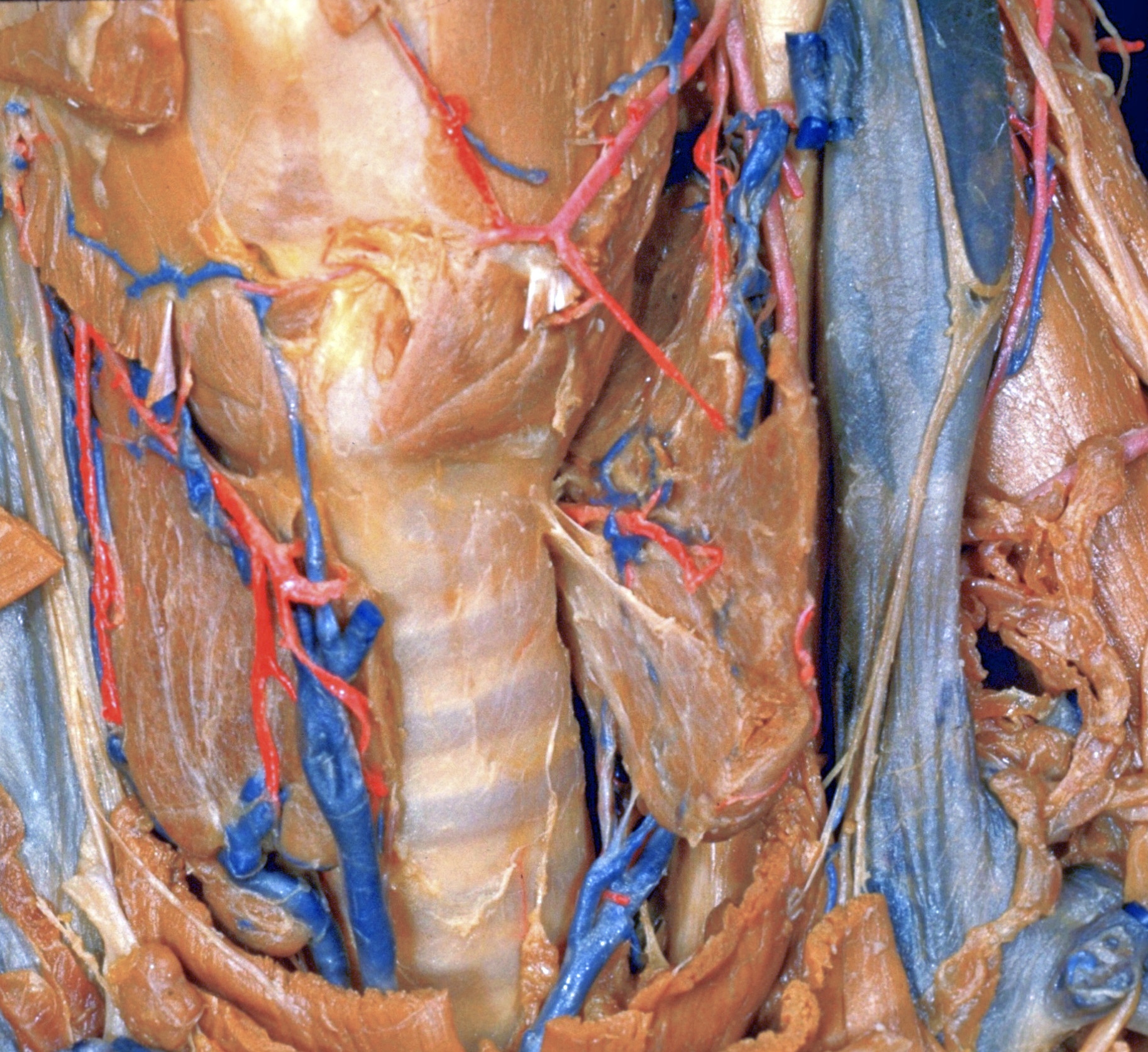
Dissection of anterior and lateral regions of neck
Relations of thyroid gland to larynx and trachea, anterior view
The isthmus (13) of the thyroid gland has been divided and the left lobe retracted laterally. The fascial attachment (9) of the gland to the upper part of the trachea is shown.
- Sternohyoid muscle right (cut across)
- Thyroid cartilage
- Thyrohyoid muscle right
- Aberrant muscular slip of cricothyroid muscle
- Prelaryngeal lymph node
- Cricothyroid muscles
- Arch of cricoid cartilage
- Branch of superior thyroid artery
- Lateral suspensory ligament of thyroid gland
- Trachea
- Right lobe of thyroid gland
- Internal jugular vein
- Isthmus of thyroid gland (divided)
- Inferior thyroid vein right and tributaries
- Sternohyoid muscle (cut across)
- Inferior thyroid vein left
- Thyrohyoid muscle
- Cricothyroid branch superior thyroid artery
- Sternothyroid muscle (cut across)
- Posterior branch superior thyroid artery
- Anterior branch superior thyroid artery
- Inferior pharyngeal constrictor muscle (m. cricopharyngeus)
- Anterior scalene muscle
- Left lobe of thyroid gland (posteromedial surface)
- Inferior thyroid artery
- Common carotid artery
- Internal jugular vein
- Phrenic nerve
- Branch from ansa hypoglossi to sternohyoid muscle
- Termination of lymphatic trunks in internal jugular vein
- Sternothyroid muscle and sternohyoid muscle