Dissection of anterior and lateral regions of neck
Relations of thyroid gland, anterior view
For permissions information regarding the use of these images,
please contact Drew Bourn at dbourn@stanford.edu
Image #74-7
Dissection of anterior and lateral regions of neck
Relations of thyroid gland, anterior view
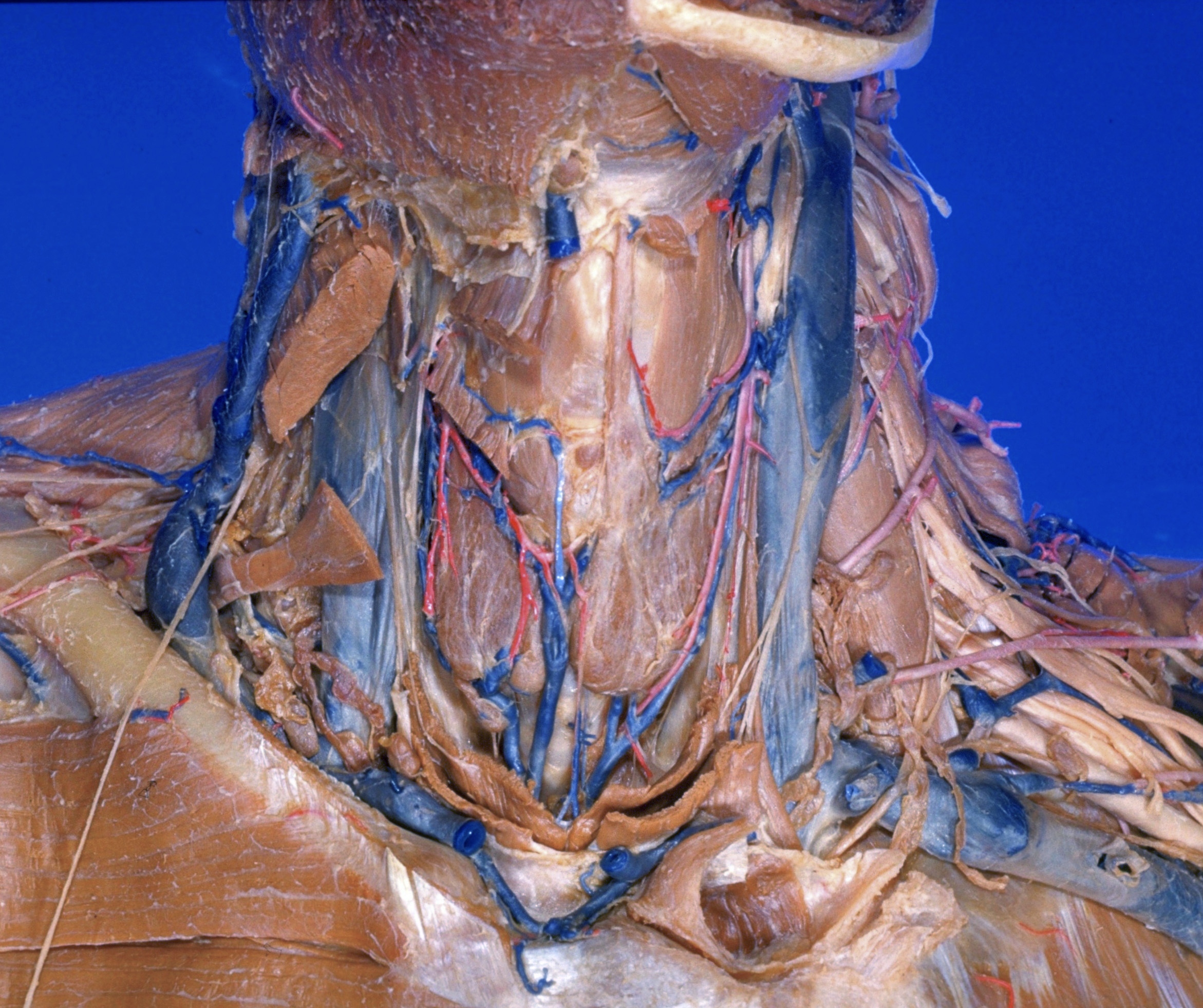
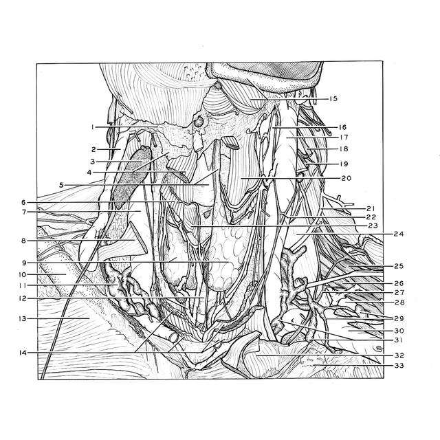
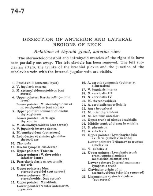
The sternocleidomastoid and infrahyoid muscles of the right side have been partially cut away. The left clavicle has been removed. The left subclavian artery, the trunks of the brachial plexus and the junction of the subclavian vein with the internal jugular vein are visible.
1
.
Superficial fascia (external layer)
2
.
External jugular vein
3
.
Sternocleidomastoid muscle (cut across)
4
.
Upper pointer: Superficial fascia (middle layer) Lower pointer: Sternohyoid muscle and omohyoid muscle (cut across)
5
.
Upper pointer: Remnant of thyroglossal duct Lower pointer: Thyroid cartilage
6
.
Sternothyroid muscle (cut across)
7
.
Internal jugular vein right
8
.
Omohyoid muscle (cut across)
9
.
Right and left lobe of thyroid gland
10
.
Clavicle
11
.
Right lymphatic duct
12
.
Upper pointer: Trachea Lower pointer: Inferior thyroid vein right