X CLOSE

Dissection of anterior and lateral regions of neck

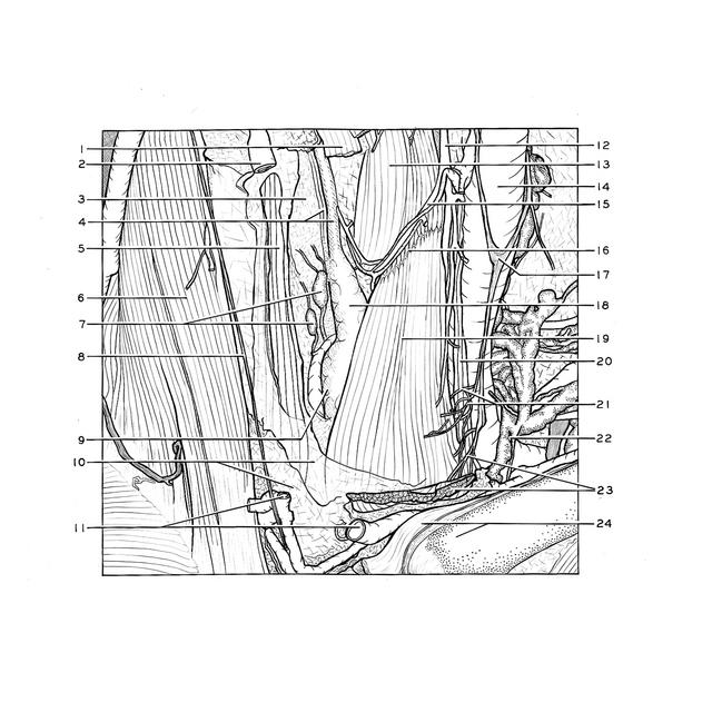
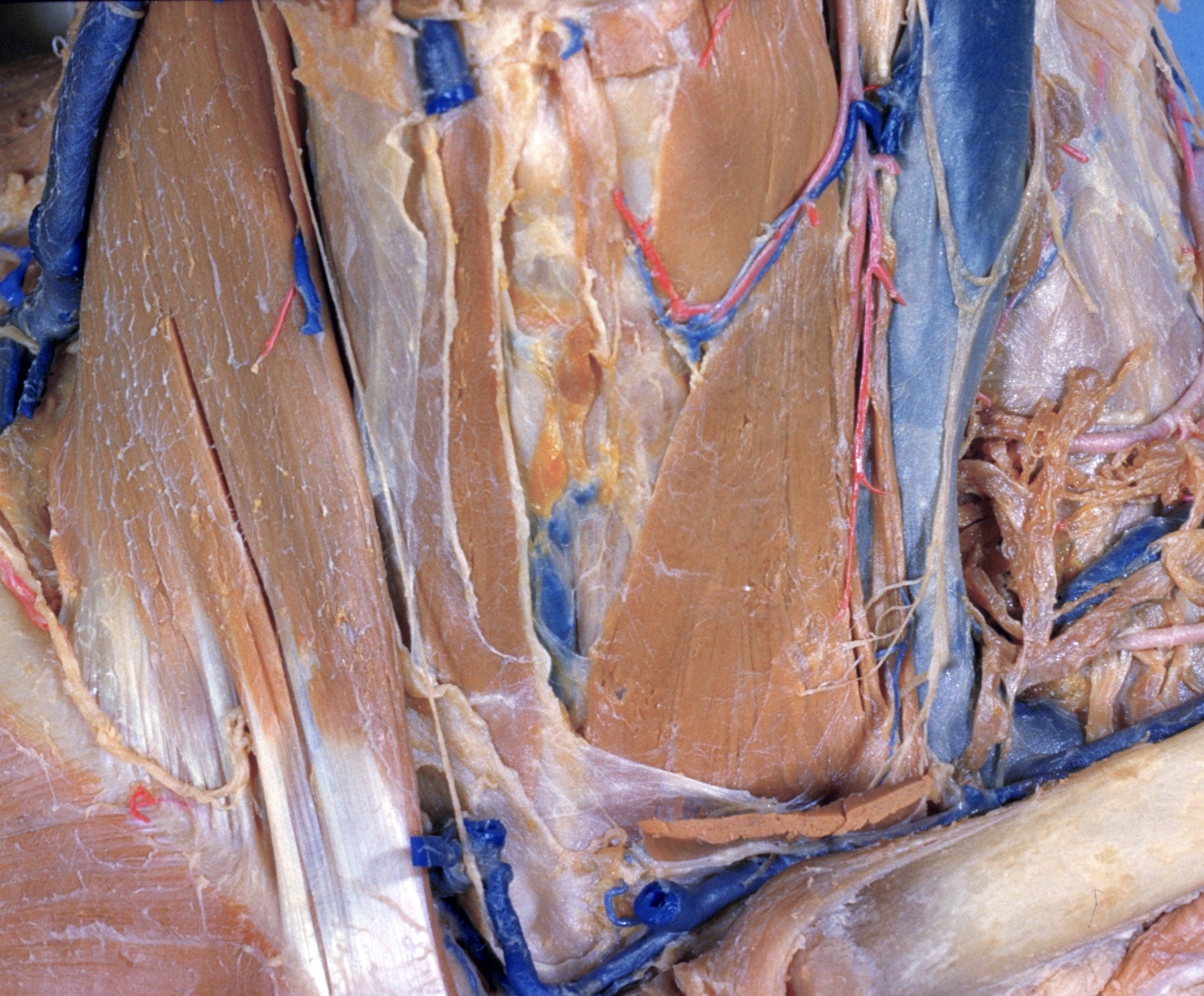
Nerve supply to sternohyoid and sternothyroid muscles, anterior view

Stanford holds the copyright to the David L. Bassett anatomical images and has assigned Creative Commons license Attribution-Share Alike 4.0 International to all of the images.
For additional information regarding use and permissions, please contact Dr. Drew Bourn at dbourn@stanford.edu.

Image #74-5
Bassett ImageDiagram Image
Legend Image
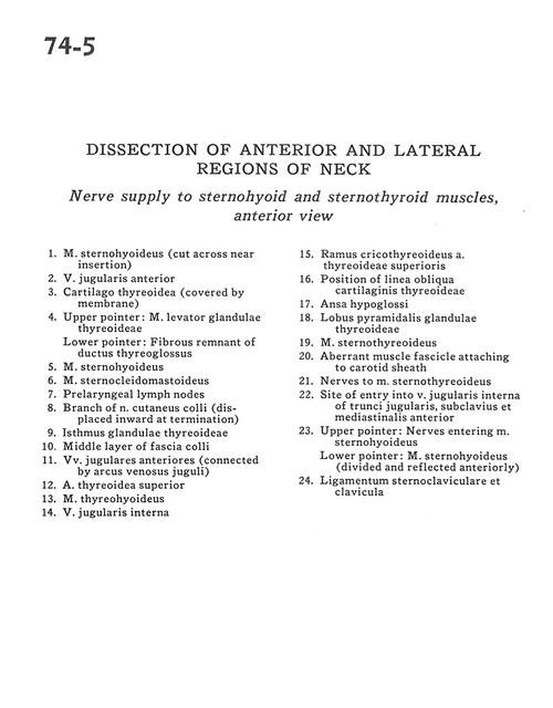
Dissection of anterior and lateral regions of neck
Nerve supply to sternohyoid and sternothyroid muscles, anterior view
1 . Sternohyoid muscle (cut across near insertion)
2 . Anterior jugular vein
3 . Thyroid cartilage (covered by membrane)
4 . Upper pointer: Levator muscle of thyroid gland Lower pointer: Fibrous-remnant of thyroglossal duct
5 . Sternohyoid muscle
6 . Sternocleidomastoid muscle
7 . Prelaryngeal lymph nodes
8 . Branch of superficial cutaneous nerve (displaced inward at termination)
9 . Isthmus of thyroid gland
10 . Middle layer of superficial fascia
11 . Anterior jugular veins (connected by jugular venous arch)
12 . Superior thyroid artery
13 . Thyrohyoid muscle
14 . Internal jugular vein
15 . Cricothyroid branch superior thyroid artery
16 . Position of oblique line thyroid cartilage
17 . Ansa hypoglossi
18 . Pyramidal lobe of thyroid gland
19 . Sternothyroid muscle
20 . Aberrant muscle fascicle attaching to carotid sheath
21 . Nerves to sternothyroid muscle
22 . Site of entry into internal jugular vein of jugular, subclavius and anterior mediastinal trunks
23 . Upper pointer: Nerves entering sternohyoid muscle Lower pointer: Sternohyoid muscle (divided and reflected anteriorly)
24 . Sternoclavicular ligament and clavicle