X CLOSE

Dissection of anterior and lateral regions of neck

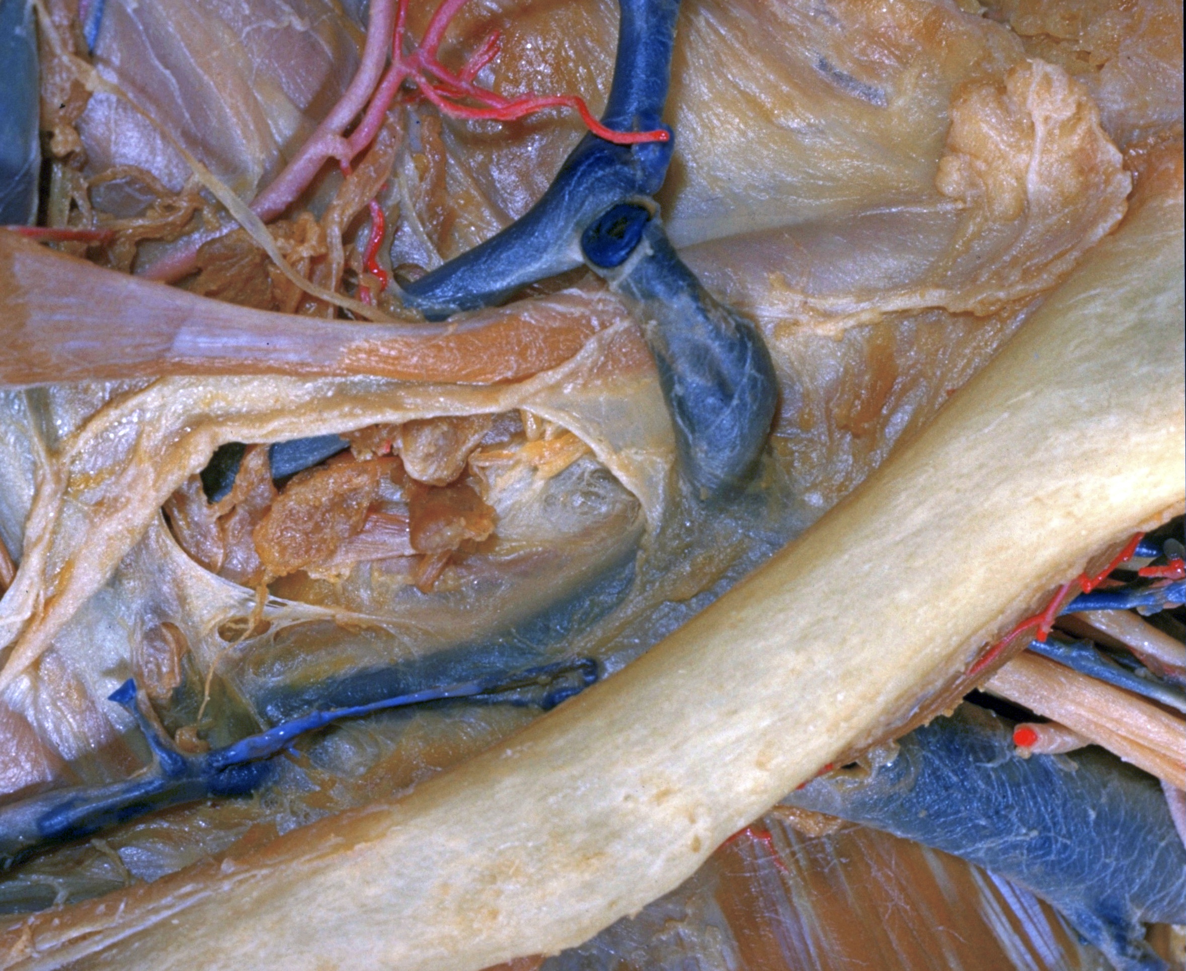
Close-up view of clavicular attachment of middle layer of cervical fascia; inferior deep cervical lymphatics, anterosuperior view

Stanford holds the copyright to the David L. Bassett anatomical images and has assigned Creative Commons license Attribution-Share Alike 4.0 International to all of the images.
For additional information regarding use and permissions, please contact Dr. Drew Bourn at dbourn@stanford.edu.

Image #74-1
Bassett ImageDiagram Image
Legend Image
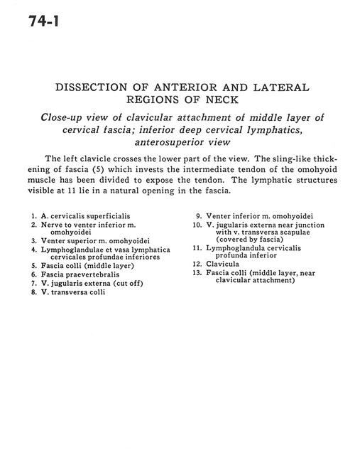
Dissection of anterior and lateral regions of neck
Close-up view of clavicular attachment of middle layer of cervical fascia; inferior deep cervical lymphatics, anterosuperior view
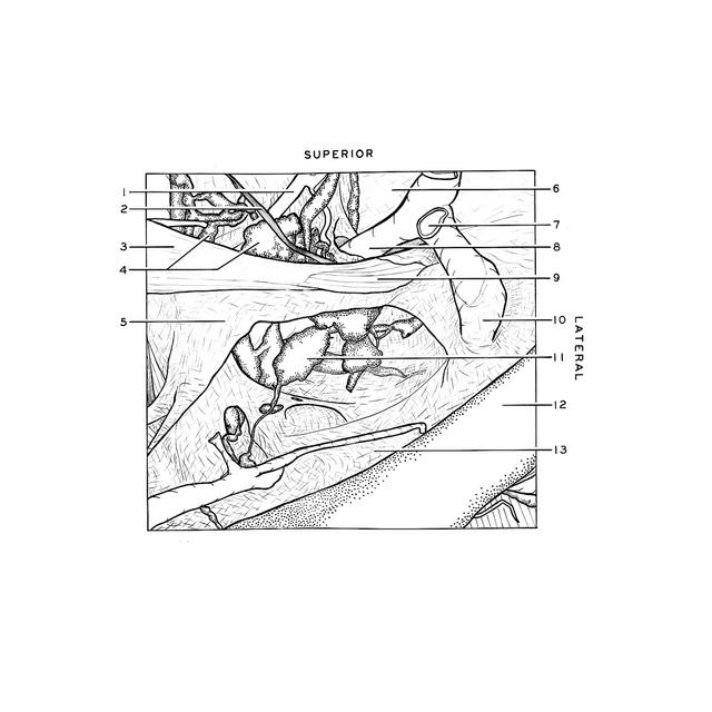
The left clavicle crosses the lower part of the view. The sling-like thickening of fascia (5) which invests the intermediate tendon of the omohyoid muscle has been divided to expose the tendon. The lympathic structures visible at 11 lie in a natural opening in the fascia.
1 . Superficial cervical artery
2 . Nerve to inferior belly omohyoid muscle
3 . Superior belly omohyoid muscle
4 . Inferior deep cervical lymph nodes and lymph vessels
5 . Superficial fascia (middle layer)
6 . Prevertebral fascia
7 . External jugular vein (cut off)
8 . Superficial transverse vein
9 . Inferior belly omohyoid muscle
10 . External jugular vein near junction with transverse scapular vein (covered by fascia)
11 . Inferior deep cervical lymph node
12 . Clavicle
13 . Superficial fascia (middle layer, near clavicular attachment)