X CLOSE

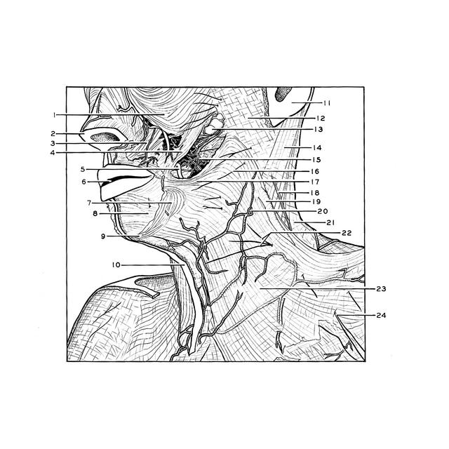
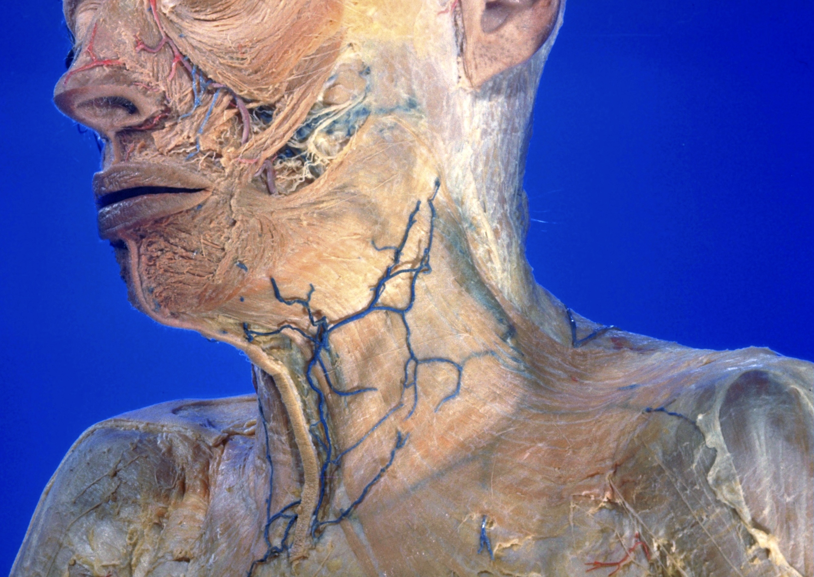
Dissection of anterior and lateral regions of neck

Superficial nerves and blood vessels; platysma, anterolateral view

Stanford holds the copyright to the David L. Bassett anatomical images and has assigned Creative Commons license Attribution-Share Alike 4.0 International to all of the images.
For additional information regarding use and permissions, please contact Dr. Drew Bourn at dbourn@stanford.edu.

Image #72-5
Bassett ImageDiagram Image
Legend Image
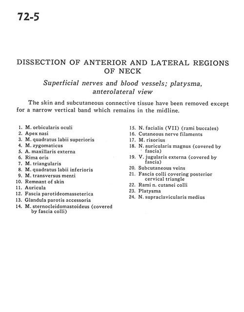
Dissection of anterior and lateral regions of neck
Superficial nerves and blood vessels; platysma, anterolateral view
The skin and subcutaneous connective tissue have been removed except for a narrow vertical band which remains in the midline.
1 . Ocular orbicular muscle
2 . Nasal apex
3 . Superior labial quadratic muscle
4 . Zygomatic muscle
5 . External maxillary artery
6 . Rima oris
7 . Triangular muscle
8 . Inferior labial quadratic muscle
9 . Transversus menti muscle
10 . Remnant of skin
11 . Auricula
12 . Parotid-masseteric fascia
13 . Accessory parotid gland
14 . Sternocleidomastoid muscle (covered by fascia colli)
15 . Facial nerve (VII) (buccal branch)
16 . Cutaneous nerve filaments
17 . Risorius muscle
18 . Auricularis magnus nerve (covered by fascia)
19 . External jugular vein (covered by fascia)
20 . Subcutaneous veins
21 . Fascia colli covering posterior cervical triangle
22 . Branch of cutaneous colli nerve
23 . Platysma
24 . Medial supraclavicular nerve
* . [Legend above restored translation from Latin]