X CLOSE

Dissection of nose

Cartilage of nasal septum

Stanford holds the copyright to the David L. Bassett anatomical images and has assigned Creative Commons license Attribution-Share Alike 4.0 International to all of the images.
For additional information regarding use and permissions, please contact Dr. Drew Bourn at dbourn@stanford.edu.

Image #71-4
Bassett ImageDiagram Image
Legend Image
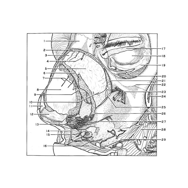
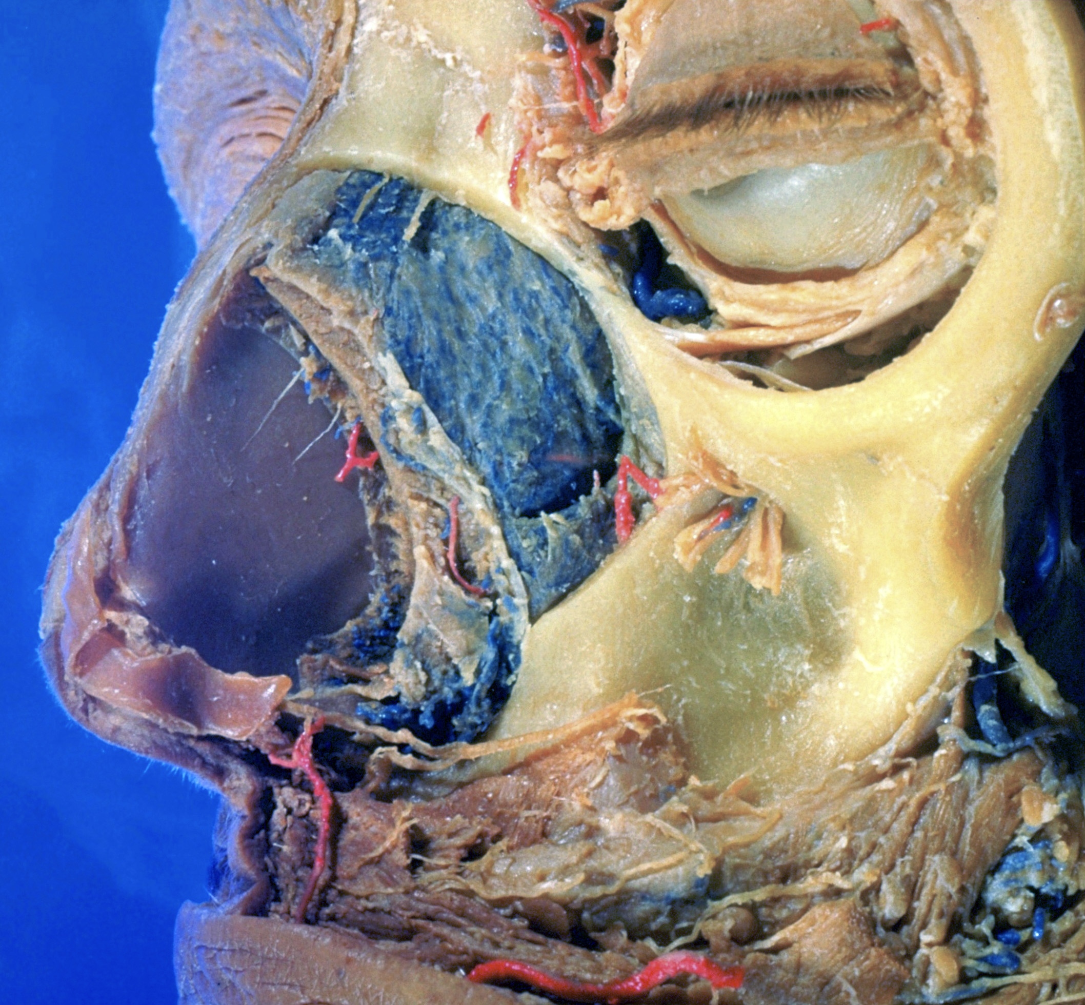
Dissection of nose
Cartilage of nasal septum
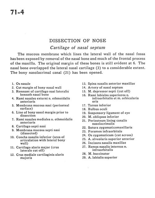
The mucous membrane which lines the lateral wall of the nasal fossa has been exposed by removal of the nasal bone and much of the frontal process of the maxilla. The original margin of these bones are still evident at 6. The nasal bone overlapped the lateral nasal cartilage (3) to a considerable extent. The bony nasolacrimal canal (21) has been opened.
1 . Nasal bone
2 . Cut margin of bony nasal wall
3 . Remnant of lateral nasal cartilage beneath nasal bone
4 . External nasal branches of anterior ethmoidal nerve
5 . Nasal mucosal membrane (periosteal surface)
6 . Line of bony nasal margin prior to dissection
7 . Middle nasal branches of anterior ethmoidal nerve
8 . Septal cartilage
9 . Mucosal membrane of nasal septum (dissected)
10 . Inferior nasal concha (area of articulation with lateral bony wall)
11 . Greater alar cartilage (lateral crus cut off)
12 . Medial crus of greater alar cartilage
13 . Anterior nasal spine maxilla
14 . Artery of nasal septum
15 . Depressor septi nasi muscle (cut off)
16 . Superior labial branches of infraorbital nerve and orbicularis oris muscle
17 . Inferior tarsus
18 . Eyeball
19 . Suspensory ligament of eye
20 . Inferior oblique muscle
21 . Periosteum lining nasolacrimal canal
22 . Zygomaticomaxillary suture
23 . Infraorbital foramen
24 . Zygomatic bone (cut across)
25 . Anterior superior alveolar artery
26 . Nasal incisure (maxilla)
27 . Internal nasal branch of infraorbital nerve
28 . Buccinator muscle
29 . Superior labial artery