X CLOSE

Oral cavity

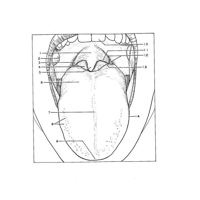
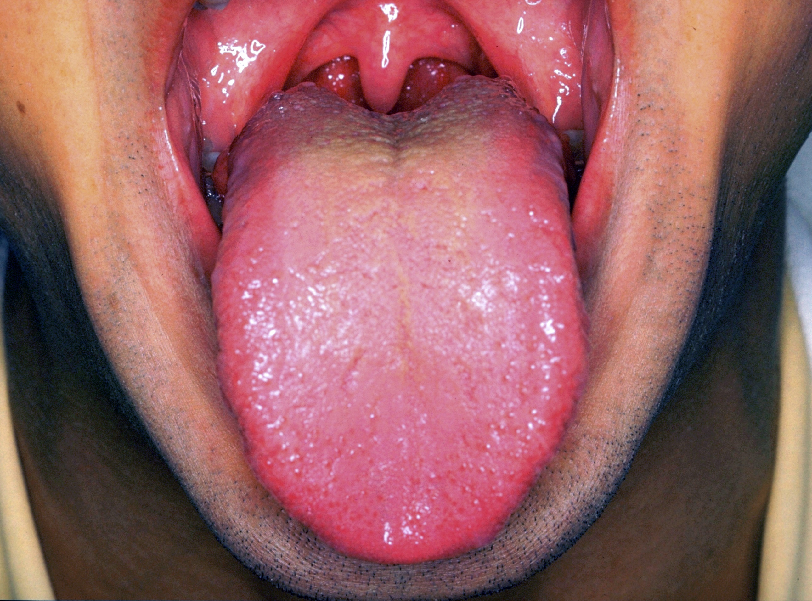
Faucial isthmus and soft palate, anterior view

Stanford holds the copyright to the David L. Bassett anatomical images and has assigned Creative Commons license Attribution-Share Alike 4.0 International to all of the images.
For additional information regarding use and permissions, please contact Dr. Drew Bourn at dbourn@stanford.edu.

Image #70-5
Bassett ImageDiagram Image
Legend Image
Oral cavity
Faucial isthmus and soft palate, anterior view
The subject is a young, adult male.
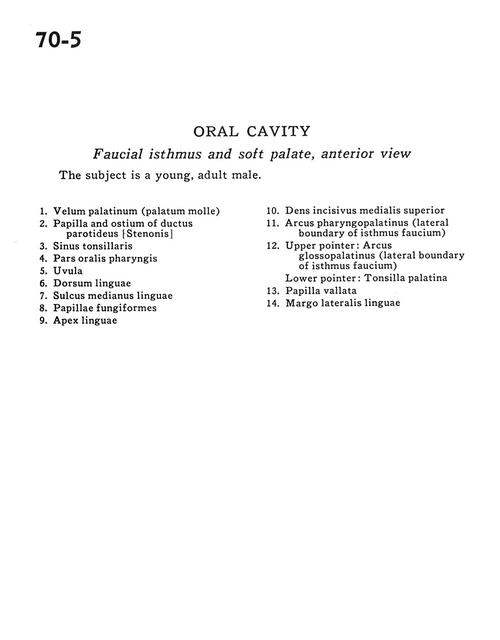
1 . Velum palatinum (soft palate)
2 . Papilla and ostium of parotid duct
3 . Tonsillar sinus
4 . Oral part pharynx
5 . Uvula
6 . Dorsum of tongue
7 . Median sulcus of tongue
8 . Fungiform papilla
9 . Apex of tongue
10 . Medial incisor (superior)
11 . Palatopharyngeal arch (lateral boundary of faucial isthmus)
12 . Upper pointer: Palatoglossal arch (lateral boundary of faucial isthmus) Lower pointer: Palatine tonsil
13 . Vallate papilla
14 . Lateral margin of tongue