X CLOSE

Dissection of oral region

Mylohyoid muscle and nerve; internal pterygoid muscle, left anterolateral view

Stanford holds the copyright to the David L. Bassett anatomical images and has assigned Creative Commons license Attribution-Share Alike 4.0 International to all of the images.
For additional information regarding use and permissions, please contact Dr. Drew Bourn at dbourn@stanford.edu.

Image #69-3
Bassett ImageDiagram Image
Legend Image
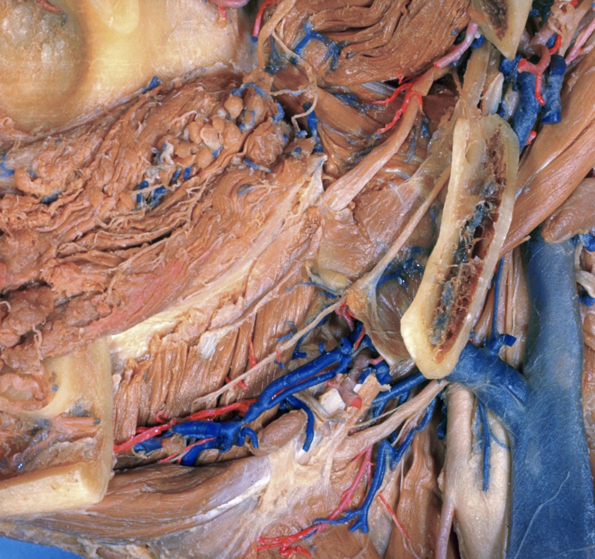
Dissection of oral region
Mylohyoid muscle and nerve; internal pterygoid muscle, left anterolateral view
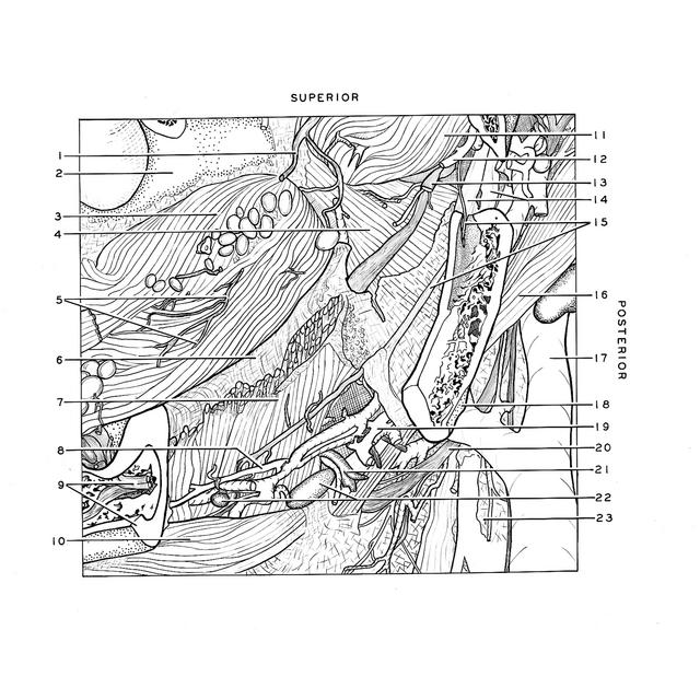
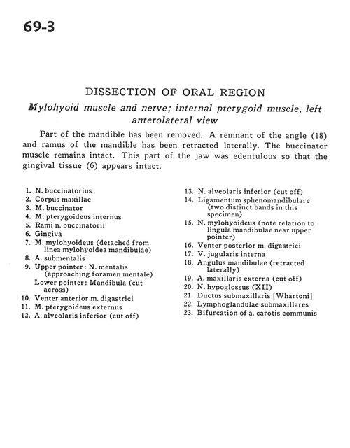
Part of the mandible has been removed. A remnant of the angle (18) and ramus of the mandible has been retracted laterally. The buccinator muscle remains intact. This part of the jaw was edentulous so that the gingival tissue (6) appears intact.
1 . Buccal nerve
2 . Body maxilla
3 . Buccinator muscle
4 . Internal pterygoid muscle
5 . Branches of buccal nerve
6 . Gingiva
7 . Mylohyoid muscle (detached from mylohyoid line mandible)
8 . Submental artery
9 . Upper pointer: Mental nerve (approaching mental foramen) Lower pointer: Mandible (cut across)
10 . Anterior belly digastric muscle
11 . External pterygoid muscle
12 . Inferior alveolar artery (cut off)
13 . Inferior alveolar nerve (cut off)
14 . Sphenomandibular ligament (two distinct bands in this specimen)
15 . Mylohyoid nerve (note relation to lingula of mandible near upper pointer)
16 . Posterior belly of digastric muscle
17 . Internal jugular vein
18 . Angle of mandible (retracted laterally)
19 . External maxillary artery (cut off)
20 . Hypoglossal nerve (XII)
21 . Submandibular duct
22 . Submandibular lymph node
23 . Bifurcation of common carotid artery