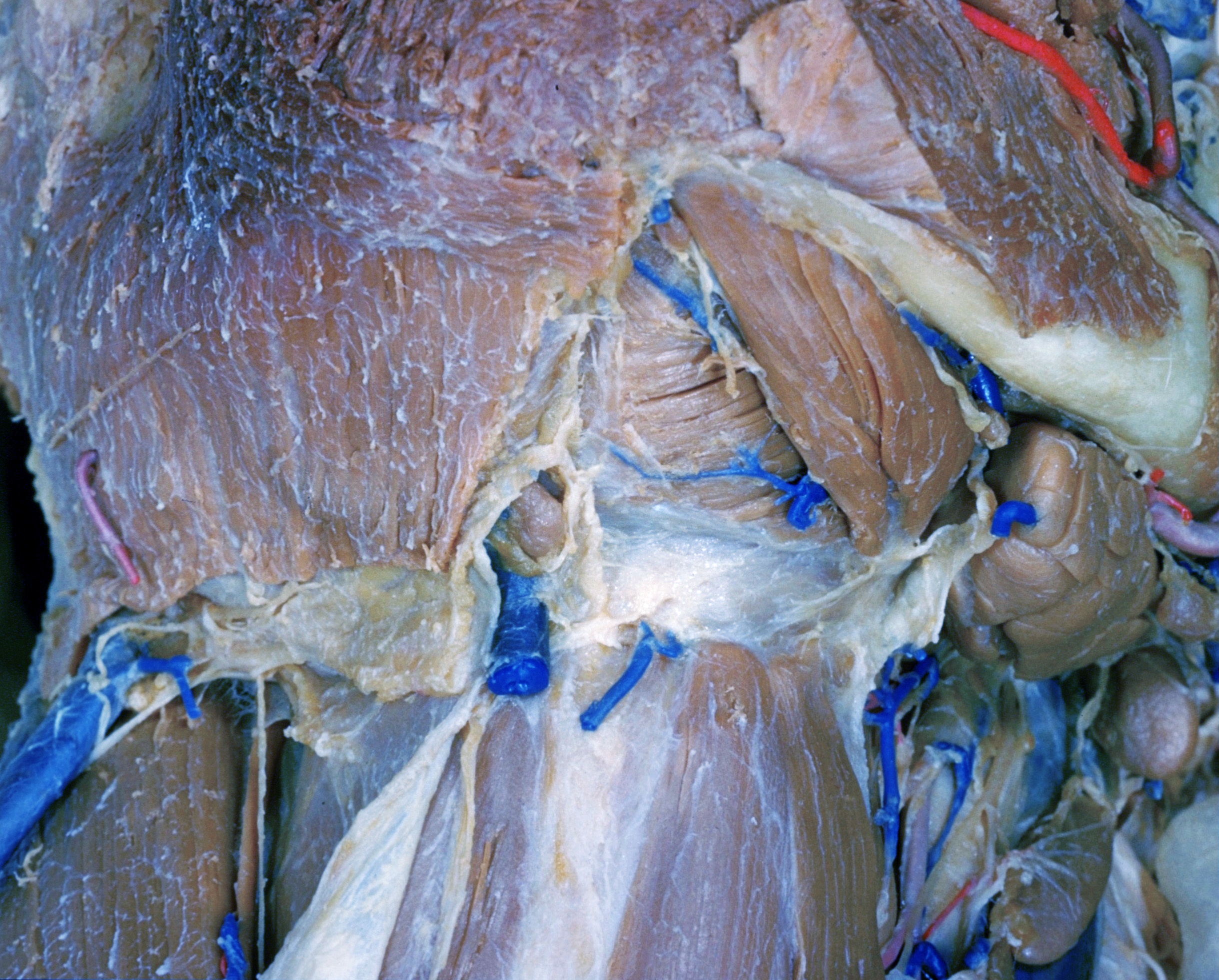
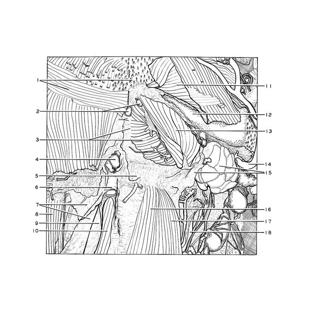
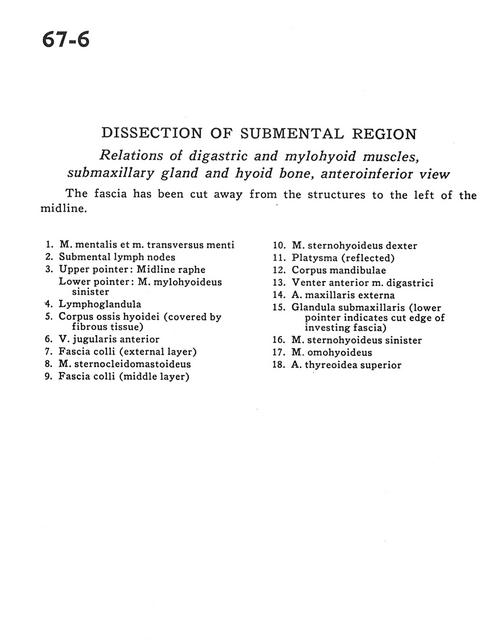
Dissection of submental region
Relations of digastric and mylohyoid muscles, submaxillary gland and hyoid bone, anteroinferior view
Stanford holds the copyright to the David L. Bassett anatomical images and has assigned
Creative Commons license Attribution-Share Alike 4.0 International to all of the images.
For additional information regarding use and permissions,
please contact Dr. Drew Bourn at dbourn@stanford.edu.
Image #67-6
 |  | ||||||||||||||||||||||||||||||||||||||||
 |
|