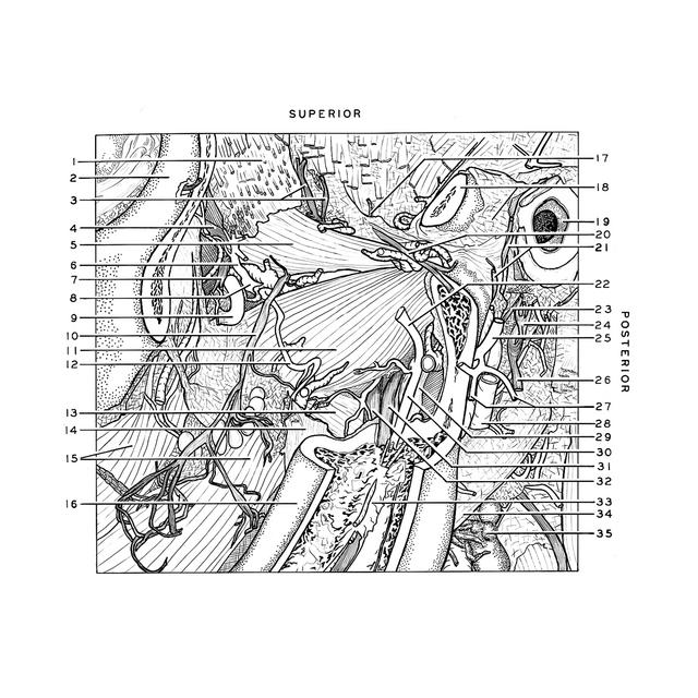
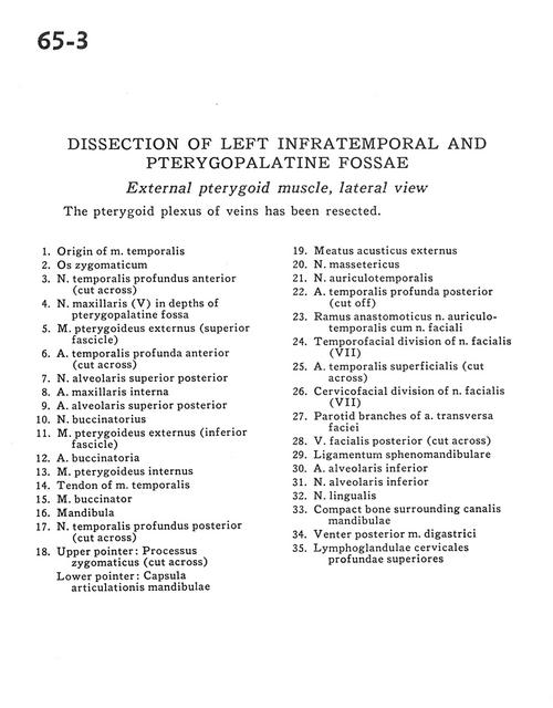
Dissection of left infratemporal and pterygopalatine fossae
External pterygoid muscle, lateral view
Stanford holds the copyright to the David L. Bassett anatomical images and has assigned
Creative Commons license Attribution-Share
Alike 4.0 International to all of the images.
For additional information regarding use and permissions,
please contact Dr. Drew Bourn at dbourn@stanford.edu.
Image #65-3
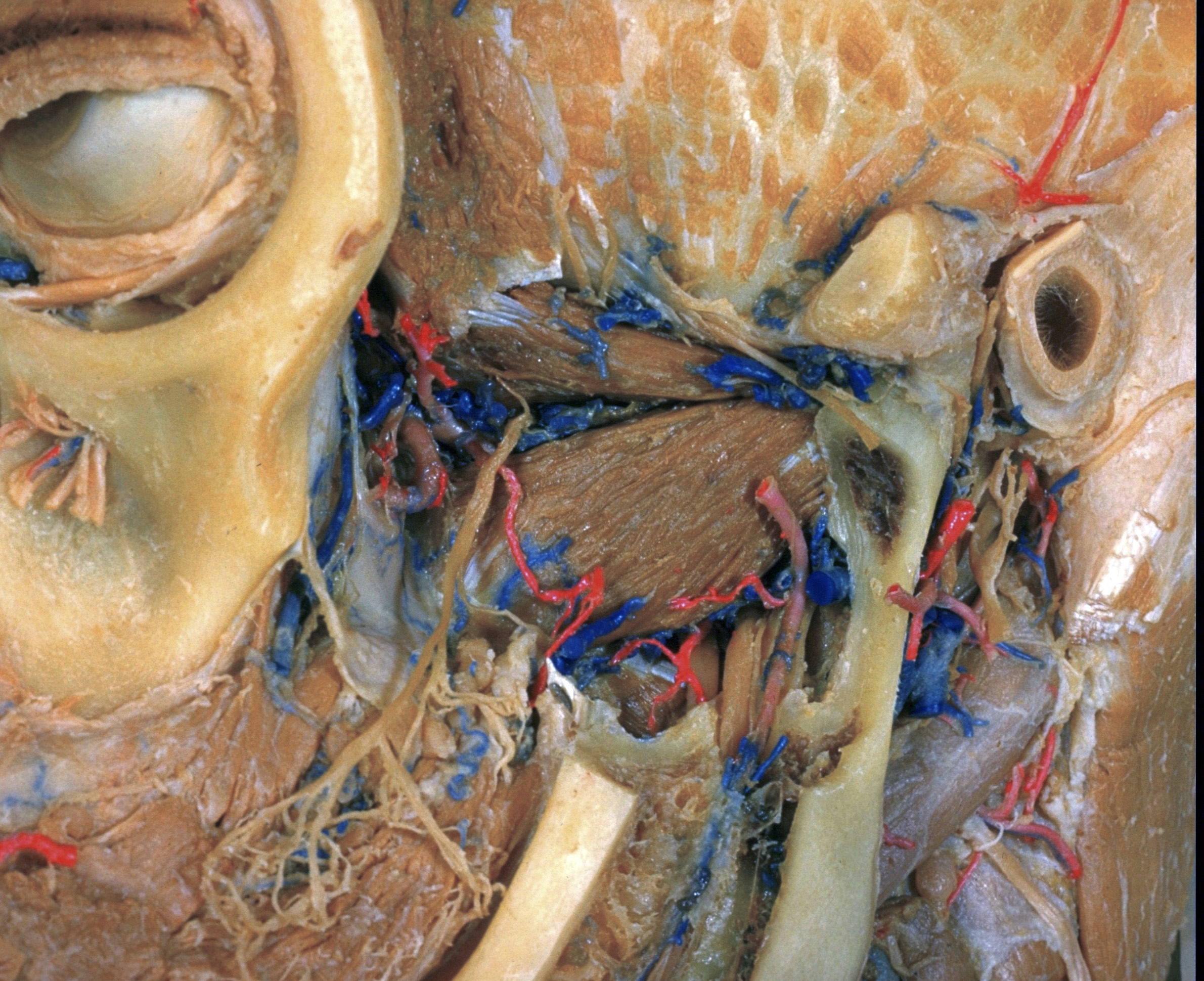
Dissection of left infratemporal and pterygopalatine fossae
External pterygoid muscle, lateral view
The pterygoid plexus of veins has been resected.
- Origin of temporalis muscle
- Zygomatic bone
- Anterior deep temporal nerve (cut across)
- Maxillary nerve (V) in depths of pterygopalatine fossa
- External pterygoid muscle (superior fascicle)
- Deep anterior temporal artery (cut across)
- Superior posterior alveolar nerve
- Internal maxillary artery
- Superior posterior alveolar artery
- Buccinator nerve
- External pterygoid muscle (inferior fascicle)
- Buccal artery
- Internal pterygoid muscle
- Tendon of temporalis muscle
- Buccinator muscle
- Mandible
- Posterior deep temporal nerve (cut across)
- Upper pointer: Zygomatic process (cut across) Lower pointer: Joint capsule of mandible
- External acoustic meatus
- Masseteric nerve
- Auriculotemporal nerve
- Deep posterior temporal artery (cut off)
- Anastomotic branch auriculotemporal nerve with facial nerve
- Temporofacial division of facial nerve (VII)
- Superficial temporal artery (cut across)
- Cervicofacial division of facial nerve (VII)
- Parotid branches of transverse facial artery
- Posterior facial vein (cut across)
- Sphenomandibular ligament
- Inferior alveolar artery
- Inferior alveolar nerve
- Lingual nerve
- Compact bone surrounding mandibular canal
- Posterior belly of digastric muscle
- Superior deep cervical lymph nodes