Dissection of left temporal region
Anterior deep temporal artery, anterolateral view
Stanford holds the copyright to the David L. Bassett anatomical images and has assigned
Creative Commons license Attribution-Share
Alike 4.0 International to all of the images.
For additional information regarding use and permissions,
please contact Dr. Drew Bourn at dbourn@stanford.edu.
Image #64-6
Dissection of left temporal region
Anterior deep temporal artery, anterolateral view
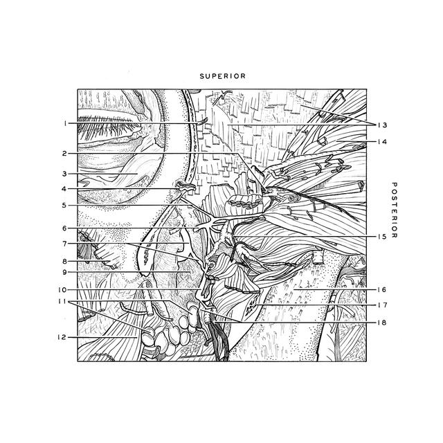
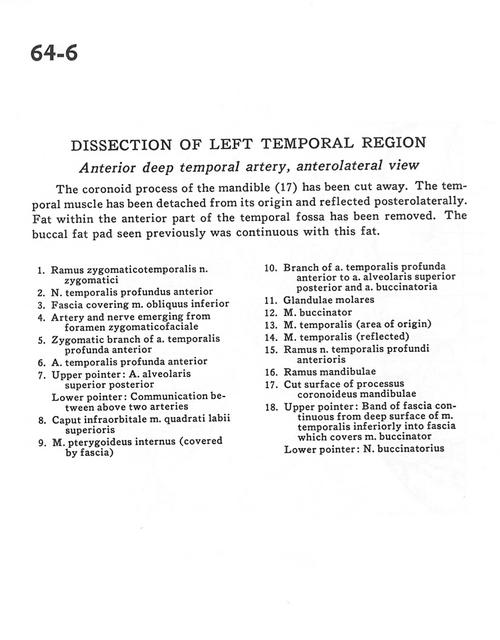
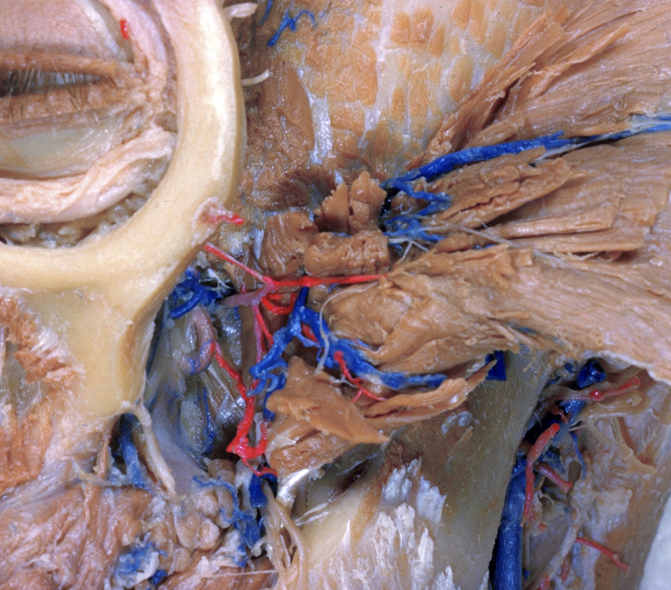
The coronoid process of the mandible (17) has been cut away. The temporal muscle has been detached from its origin and reflected posterolaterally. Fat within the anterior part of the temporal fossa has been removed. The buccal fat pad seen previously was continuous with this fat.
- Zygomaticotemporal branch zygomatic nerve
- Anterior deep temporal nerve
- Fascia covering inferior oblique muscle
- Artery and nerve emerging from zygomaticofacial foramen
- Zygomatic branch of deep anterior temporal artery
- Deep anterior temporal artery
- Upper pointer: Superior posterior alveolar artery Lower pointer: Communication between above two arteries
- Infraorbital head of levator labii superioris muscle
- Internal pterygoid muscle (covered by fascia)
- Branch of deep anterior temporal artery to superior posterior alveolar artery and buccal artery
- Molar glands
- Buccinator muscle
- Temporalis muscle (area of origin)
- Temporalis muscle (reflected)
- Branch anterior deep temporal nerve
- Ramus of mandible
- Cut surface of coronoid process of mandible
- Upper pointer: Band of fascia continuous from deep surface of temporalis muscle inferiorly into fascia which covers buccinator muscle Lower pointer: Buccal nerve