Dissection of left temporal region
Deep relations of temporal fascia, lateral view
Stanford holds the copyright to the David L. Bassett anatomical images and has assigned
Creative Commons license Attribution-Share
Alike 4.0 International to all of the images.
For additional information regarding use and permissions,
please contact Dr. Drew Bourn at dbourn@stanford.edu.
Image #64-3
Dissection of left temporal region
Deep relations of temporal fascia, lateral view
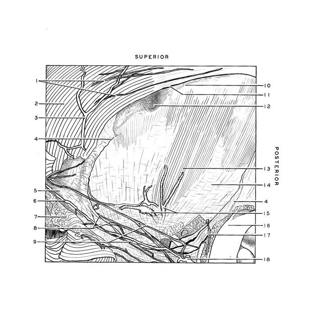
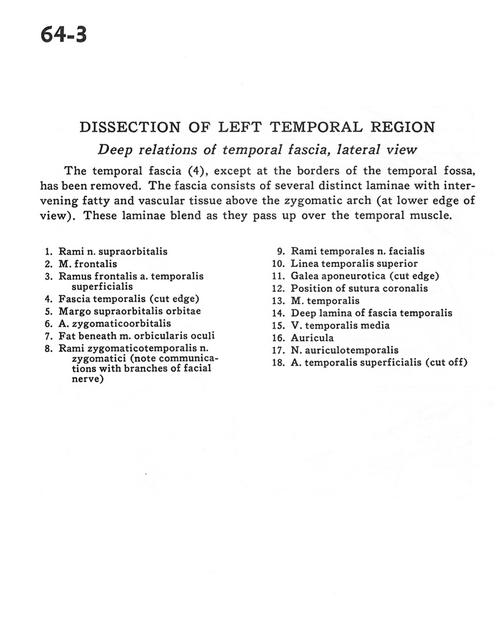
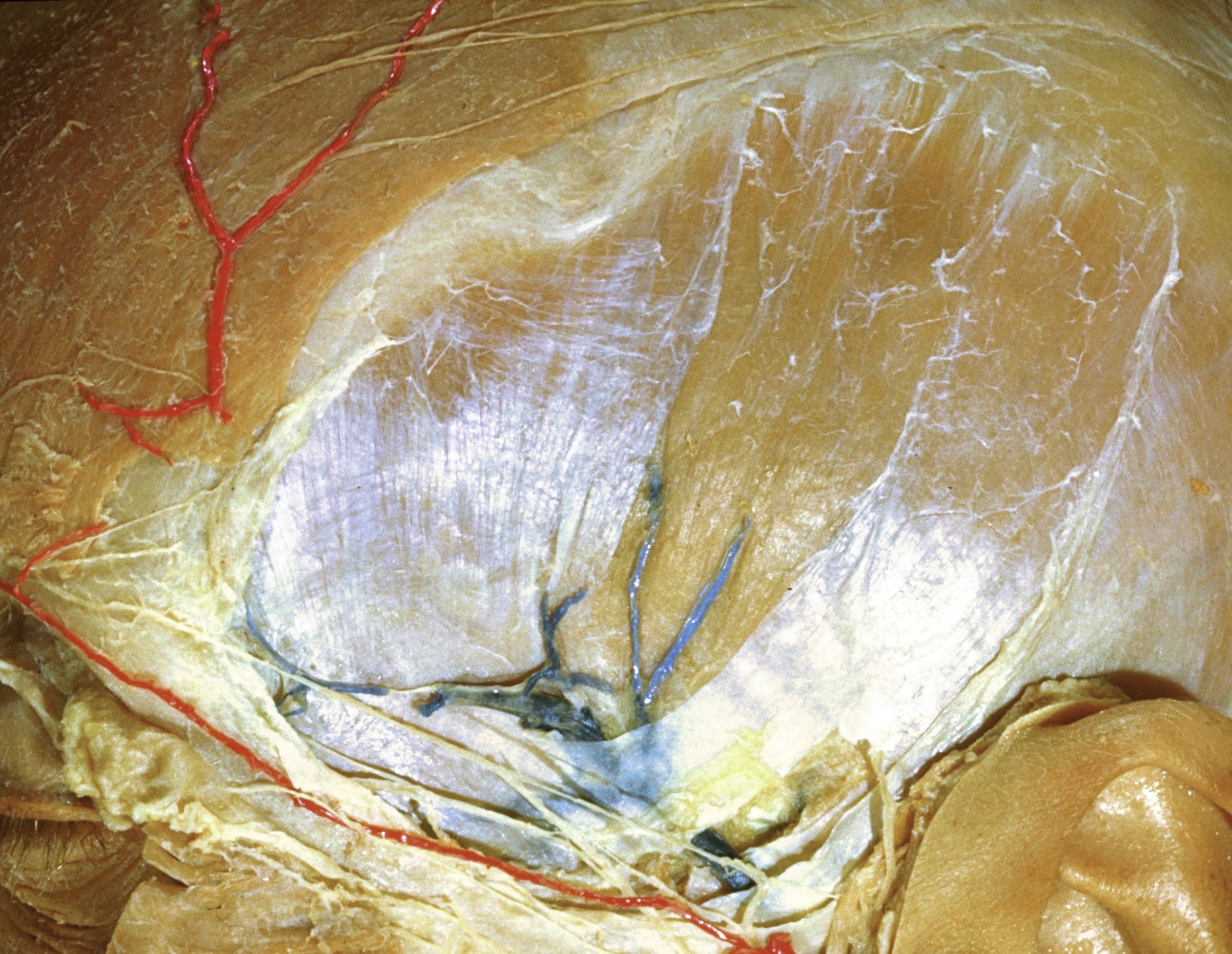
The temporal fascia (4), except at the borders of the temporal fossa, has been removed. The fascia consists of several distinct laminae with intervening fatty and vascular tissue above the zygomatic arch (at lower edge of view). These laminae blend as they pass up over the temporal muscle.
- Branches of supraorbital nerve
- Frontalis muscle
- Frontal branch of superficial temporal artery
- Temporal fascia (cut edge)
- Supraorbital margin
- Zygomatico-orbital artery
- Fat beneath orbicularis oculi muscle
- Zygomaticotemporal branches of zygomatic nerve (note communications with branches of facial nerve)
- Temporal branches of facial nerve
- Superior temporal line
- Galea aponeurotica (cut edge)
- Position of coronal suture
- Temporalis muscle
- Deep lamina of temporal fascia
- Middle temporal vein
- Auricle
- Auriculotemporal nerve
- Superficial temporal artery (cut off)