X CLOSE

Dissection of left buccal region

Buccinator muscle; buccal and molar glands; parotid duct; lateral view

Stanford holds the copyright to the David L. Bassett anatomical images and has assigned Creative Commons license Attribution-Share Alike 4.0 International to all of the images.
For additional information regarding use and permissions, please contact Dr. Drew Bourn at dbourn@stanford.edu.

Image #63-7
Bassett ImageDiagram Image
Legend Image
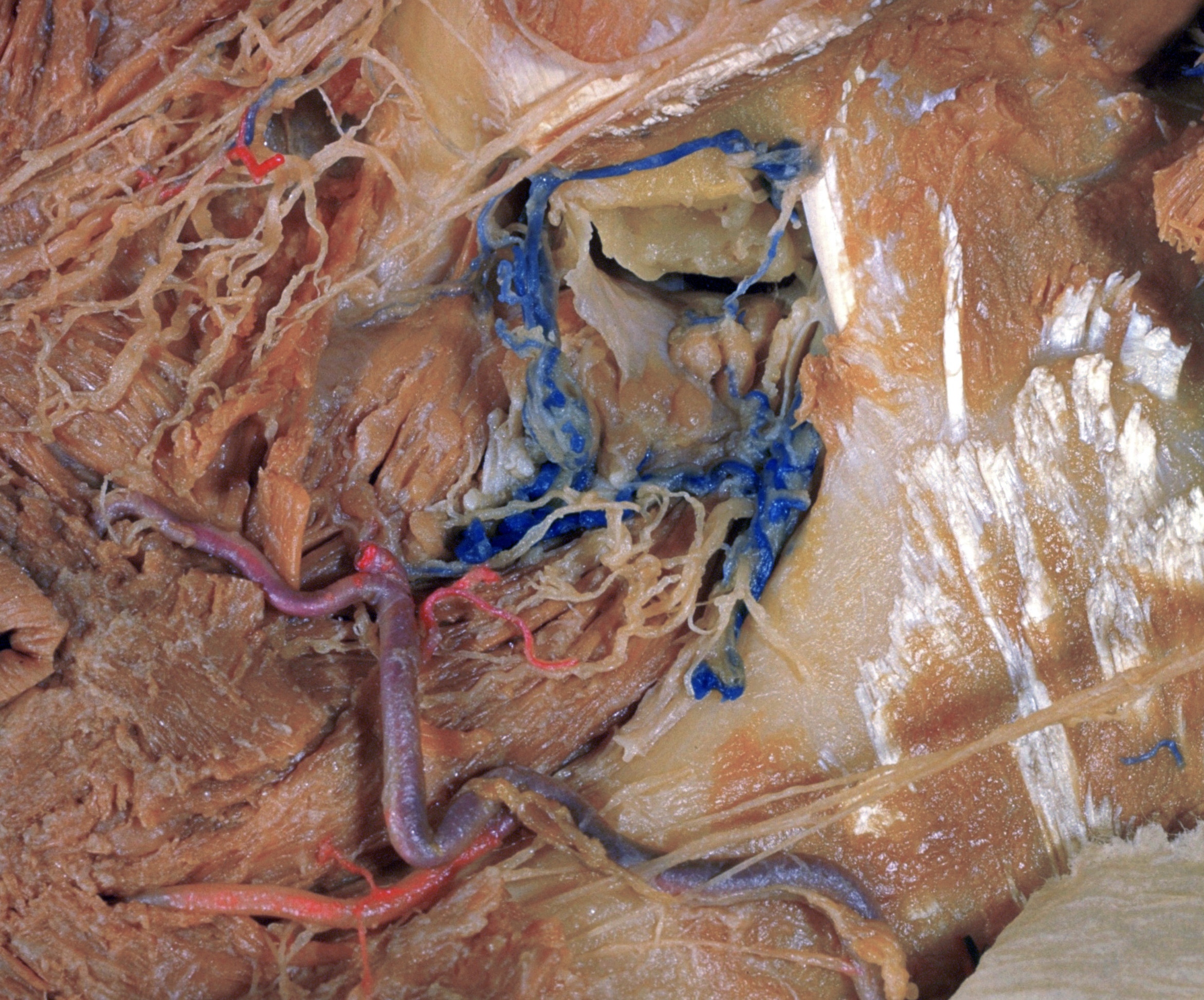
Dissection of left buccal region
Buccinator muscle; buccal and molar glands; parotid duct; lateral view
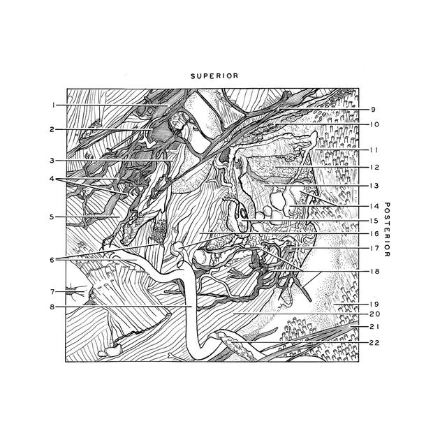
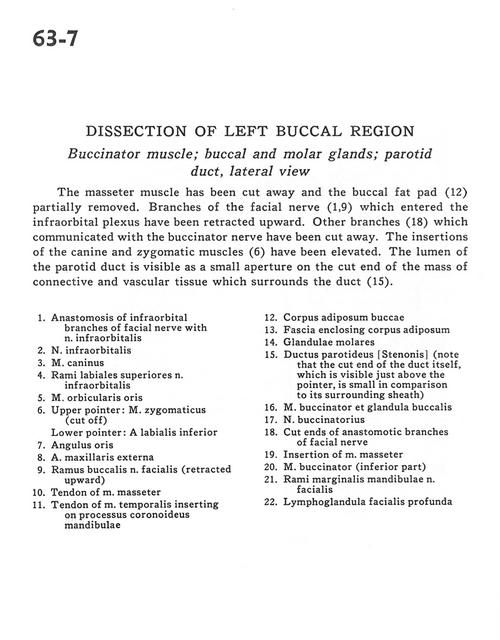
The masseter muscle has been cut away and the buccal fat pad (12) partially removed. Branches of the facial nerve (1,9) which entered the infraorbital plexus have been retracted upward. Other branches (18) which communicated with the buccinator nerve have been cut away. The insertions of canine and zygomatic muscles (6) have been elevated. The lumen of the parotid duct is visible as a small aperture on the cut end of the mass of connective and vascular tissue which surrounds the duct(15).
1 . Anastomosis of infraorbital branches of facial nerve with infraorbital nerve
2 . Infraorbital nerve
3 . Depressor anguli oris muscle
4 . Superior labial branches infraorbital nerve
5 . Orbicularis oris muscle
6 . Upper pointer: Zygomaticus muscle (cut off) Lower pointer: Inferior labial artery
7 . Angle of the mouth
8 . External maxillary artery
9 . Buccal branch facial nerve (retracted upward)
10 . Tendon of masseter muscle
11 . Tendon of temporalis muscle inserting on coronoid process of mandible
12 . Buccal fat pad
13 . Fascia enclosing fat pad
14 . Molar glands
15 . Parotid duct (note that the cut end of the duct itself, which is visible just above the pointer, is small in comparison to its surrounding sheath)
16 . Buccinator muscle and buccal glands
17 . Buccal nerve
18 . Cut ends of anastomotic branches of facial nerve
19 . Insertion of masseter muscle
20 . Buccinator muscle (inferior part)
21 . Marginal mandibular branches facial nerve
22 . Deep facial lymph node