X CLOSE

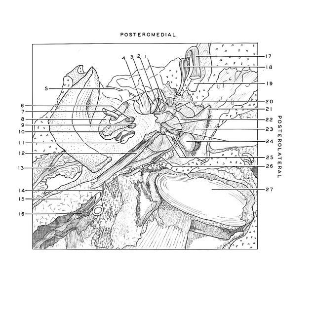
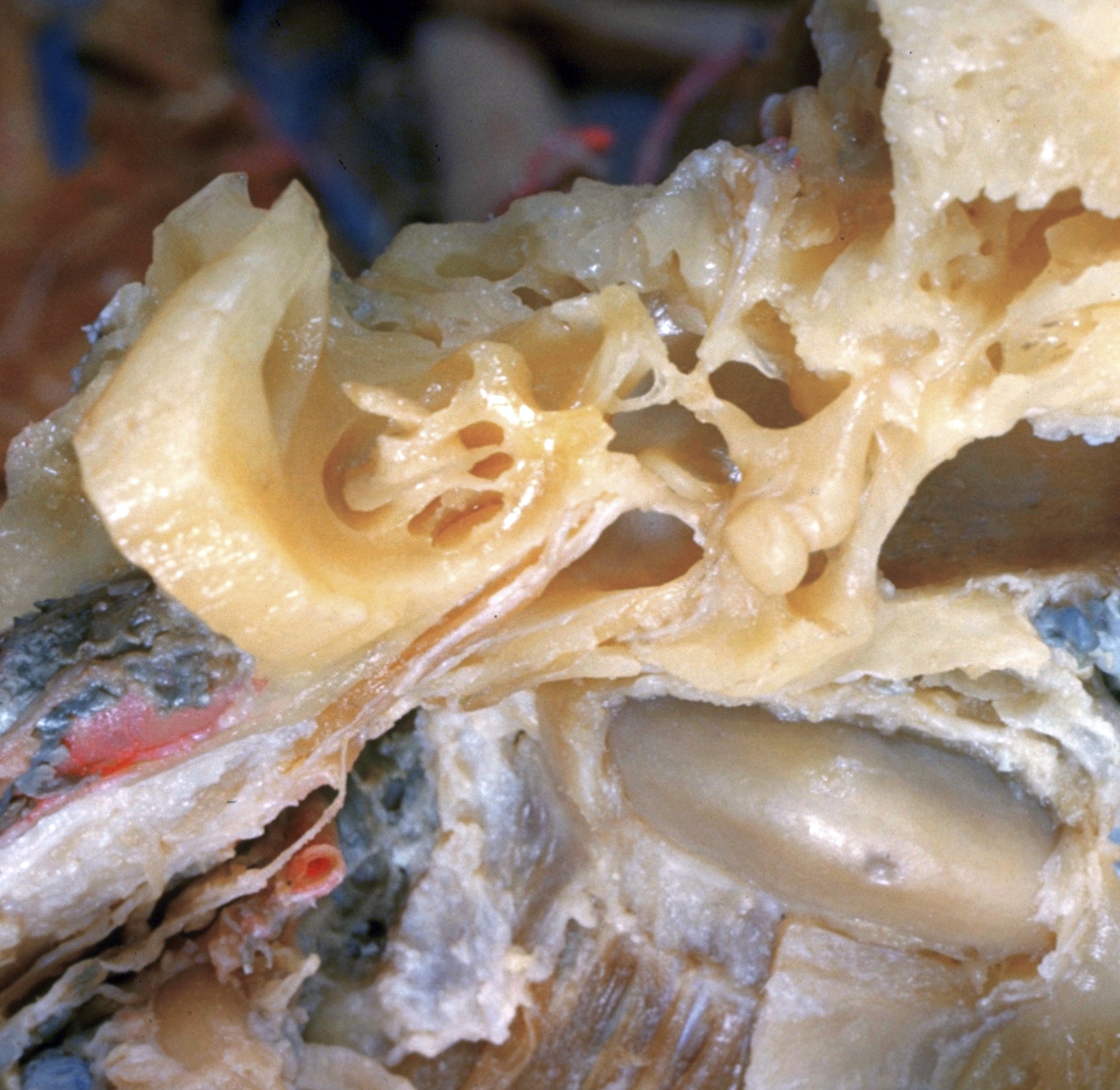
Dissection of left ear from superior aspect

Cochlea; ear ossicles in situ, anterosuperior view

Stanford holds the copyright to the David L. Bassett anatomical images and has assigned Creative Commons license Attribution-Share Alike 4.0 International to all of the images.
For additional information regarding use and permissions, please contact Dr. Drew Bourn at dbourn@stanford.edu.

Image #62-1
Bassett ImageDiagram Image
Legend Image
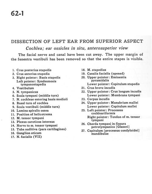
Dissection of left ear from superior aspect
Cochlea; ear ossicles in situ, anterosuperior view
The facial nerve and canal have been cut away. The upper margin of the fenestra vestibuli has been removed so that the entire staples is visible.
1 . Posterior crus of stapes
2 . Anterior crus of stapes
3 . Right pointer: Base of stapes Left pointer: Syndesmosis tympanostapedia
4 . Vestibule
5 . Tympanic nerve
6 . Scala tympani (middle turn)
7 . Vestibulocochlear nerve (VIII) (cochlear part) entering base of modiolus
8 . Basal turn of cochlea
9 . Scala vestibuli (middle turn)
10 . Osseous spiral lamina
11 . Position of helicotrema
12 . Tensor tympani muscle
13 . Internal carotid plexus
14 . Nerve to tensor tympani muscle
15 . Auditory tube (cartilaginous part)
16 . Otic ganglion
17 . Facial nerve (VII)
18 . Stapedius muscle
19 . Facial canal (opened)
20 . Upper pointer: Pyramidal eminence Lower pointer: Capitulum of stapes
21 . Short crus of incus
22 . Upper pointer: Long crus of incus Lower pointer: Tympanic membrane
23 . Body of incus
24 . Upper pointer: Manubrium of malleus Lower pointer: Capitulum of malleus
25 . Left pointer: Cochleariform process Right pointer: Tendon of tensor tympani muscle
26 . Chorda tympani in petrotympanic fissure
27 . Capitulum (condyloid process) of mandible