Dissection of left ear from superior aspect
Tympanic membrane
Stanford holds the copyright to the David L. Bassett anatomical images and has assigned
Creative Commons license Attribution-Share
Alike 4.0 International to all of the images.
For additional information regarding use and permissions,
please contact Dr. Drew Bourn at dbourn@stanford.edu.
Image #61-7
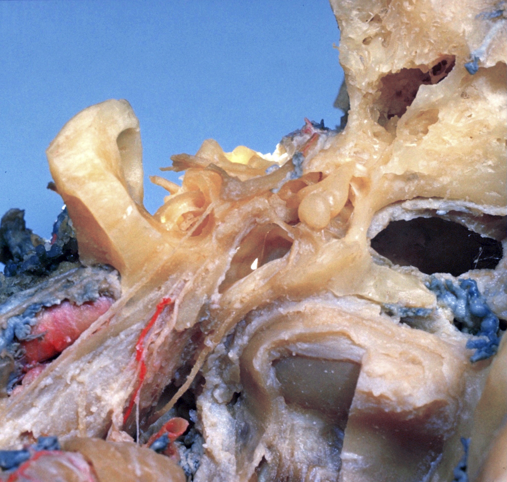
Dissection of left ear from superior aspect
Tympanic membrane
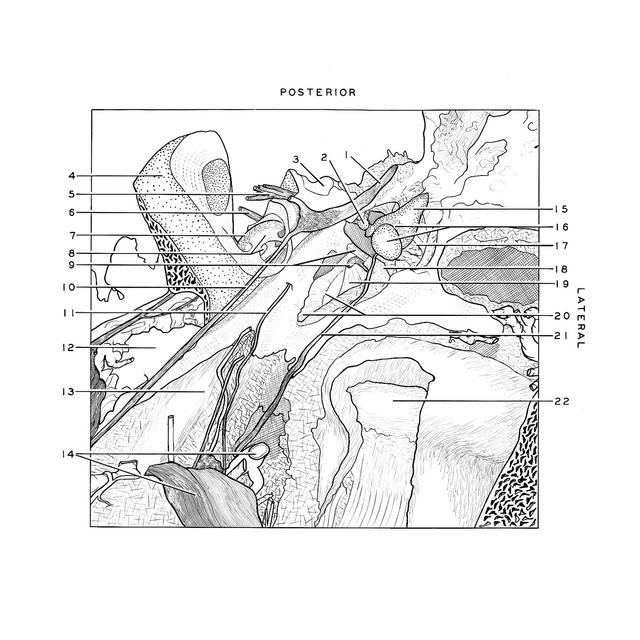
- Facial nerve (VII)
- Capitulum of stapes
- Vestibule (opened)
- Superior pyramidal angle
- Vestibulocochlear nerve (vestibular part)
- Vestibulocochlear nerve (cochlear part)
- Osseous spiral lamina
- Modiolus: Upper pointer indicates scala vestibuli of middle turn; lower pointer indicates scala tympani of apical turn)
- Upper pointer: Tendon of tensor tympani muscle Lower pointer: Manubrium of malleus
- Major superficial petrosal nerve
- Minor superficial petrosal nerve
- Internal carotid artery within carotid canal
- Outer surface of auditory tube (Eustachian)
- Middle meningeal artery and mandibular nerve
- Incus
- Upper pointer: Incus-malleolar articulation Lower pointer: Malleus
- Flaccid part of tympanic membrane
- Anterior malleolar fold
- Pars tensa tympanic membrane
- Upper pointer: Limbus (border) of tympanic membrane Lower pointer: Jugular walls of tympanum
- Chorda tympani
- Capitulum condyloid process of mandible