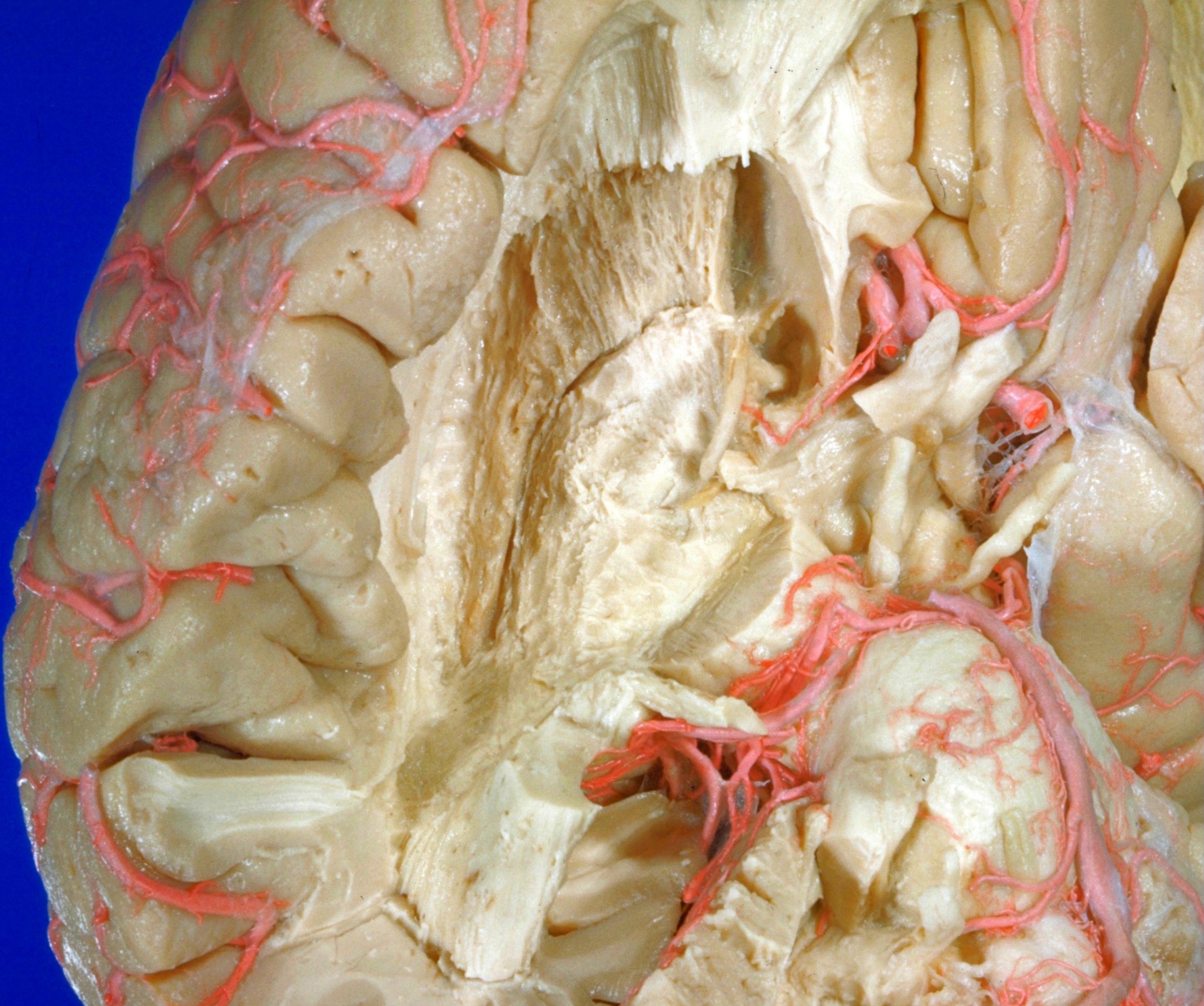
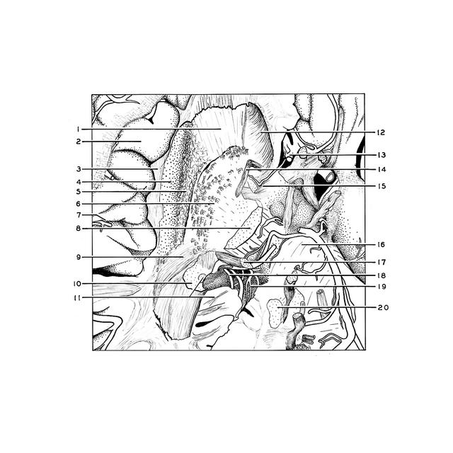
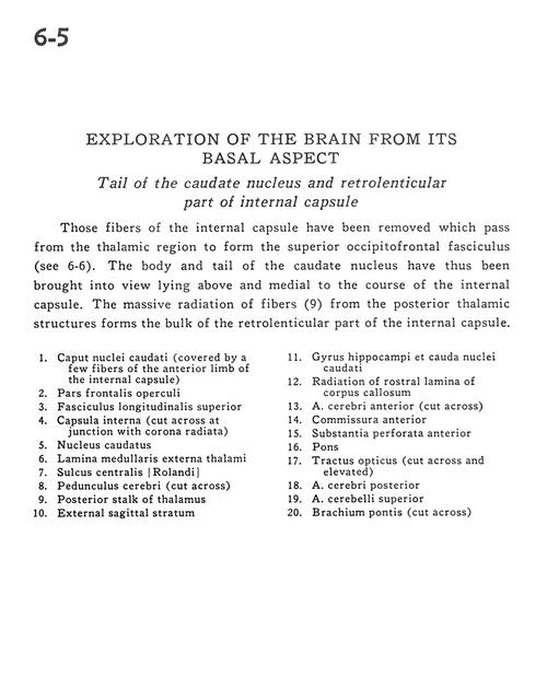
Tail of the caudate nucleus and retrolenticular part of the internal capsule
For permissions information regarding the use of these images,
please contact Drew Bourn at dbourn@stanford.edu
Image #6-5
Exploration of the brain from its basal aspect
Tail of the caudate nucleus and retrolenticular part of the internal capsule
Those fibers of the internal capsule have been removed which pass from the thalamic region to form the superior occipitofrontal fasciculus (see 6-6). The body and tail of the caudate nucleus have thus been brought into view lying above and medial to the course of the internal capsule. The massive radiation of fibers (9) from the posterior thalamic structures forms the bulk of the retrolenticular part of the internal capsule.
1
.
Head of caudate nucleus (covered by a few fibers of the anterior limb of the internal capsule)
2
.
Frontal part of operculum
3
.
Superior longitudinal fasciculus
4
.
Internal capsule (cut across at junction with corona radiata)