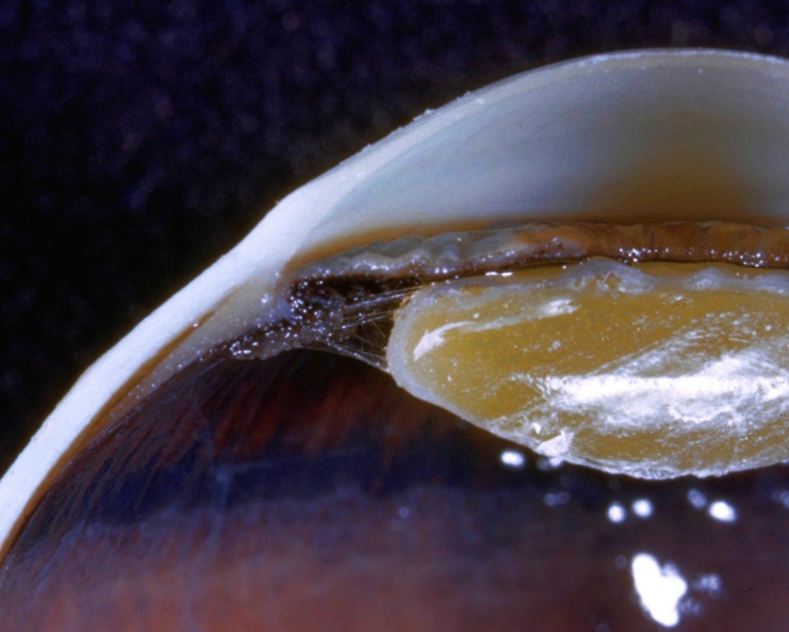
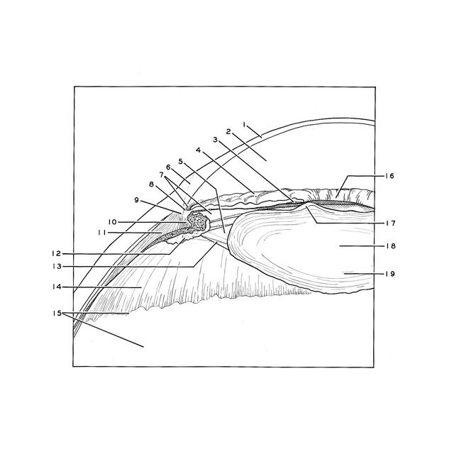
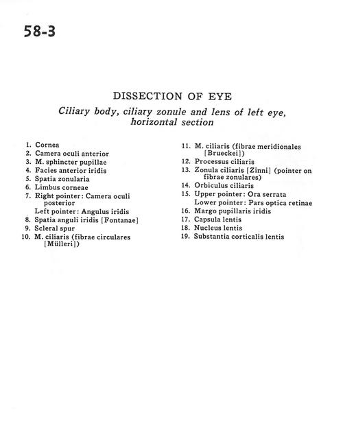
X CLOSE

Dissection of eye

Ciliary body, ciliary zonule and lens of the left eye, horizontal section

Stanford holds the copyright to the David L. Bassett anatomical images and has assigned Creative Commons license Attribution-Share Alike 4.0 International to all of the images.
For additional information regarding use and permissions, please contact Dr. Drew Bourn at dbourn@stanford.edu.

Image #58-3
Bassett ImageDiagram Image
Legend Image
Dissection of eye
Ciliary body, ciliary zonule and lens of the left eye, horizontal section
1 . Cornea
2 . Anterior chamber of eye
3 . Pupillary sphincter muscle
4 . Anterior surface of iris
5 . Zonular space
6 . Limbus of cornea
7 . Right pointer: Posterior chamber of eye Left pointer: Angle of iris
8 . Angular space of iris
9 . Scleral spur
10 . Ciliary muscle (circular fibers)
11 . Ciliary muscle (circular fibers)
12 . Ciliary process
13 . Zonula ciliaris (pointer on zonular fibers)
14 . Orbiculus ciliaris
15 . Upper pointer: Ora serrata Lower pointer: Optic (visual) part of retina
16 . Iris-pupillary margin
17 . Capsule of lens
18 . Nucleus of lens
19 . Substantia corticalis of lens