Dissection of eye
Interior of left eye cut in frontal plane
Stanford holds the copyright to the David L. Bassett anatomical images and has assigned
Creative Commons license Attribution-Share
Alike 4.0 International to all of the images.
For additional information regarding use and permissions,
please contact Dr. Drew Bourn at dbourn@stanford.edu.
Image #58-1
Dissection of eye
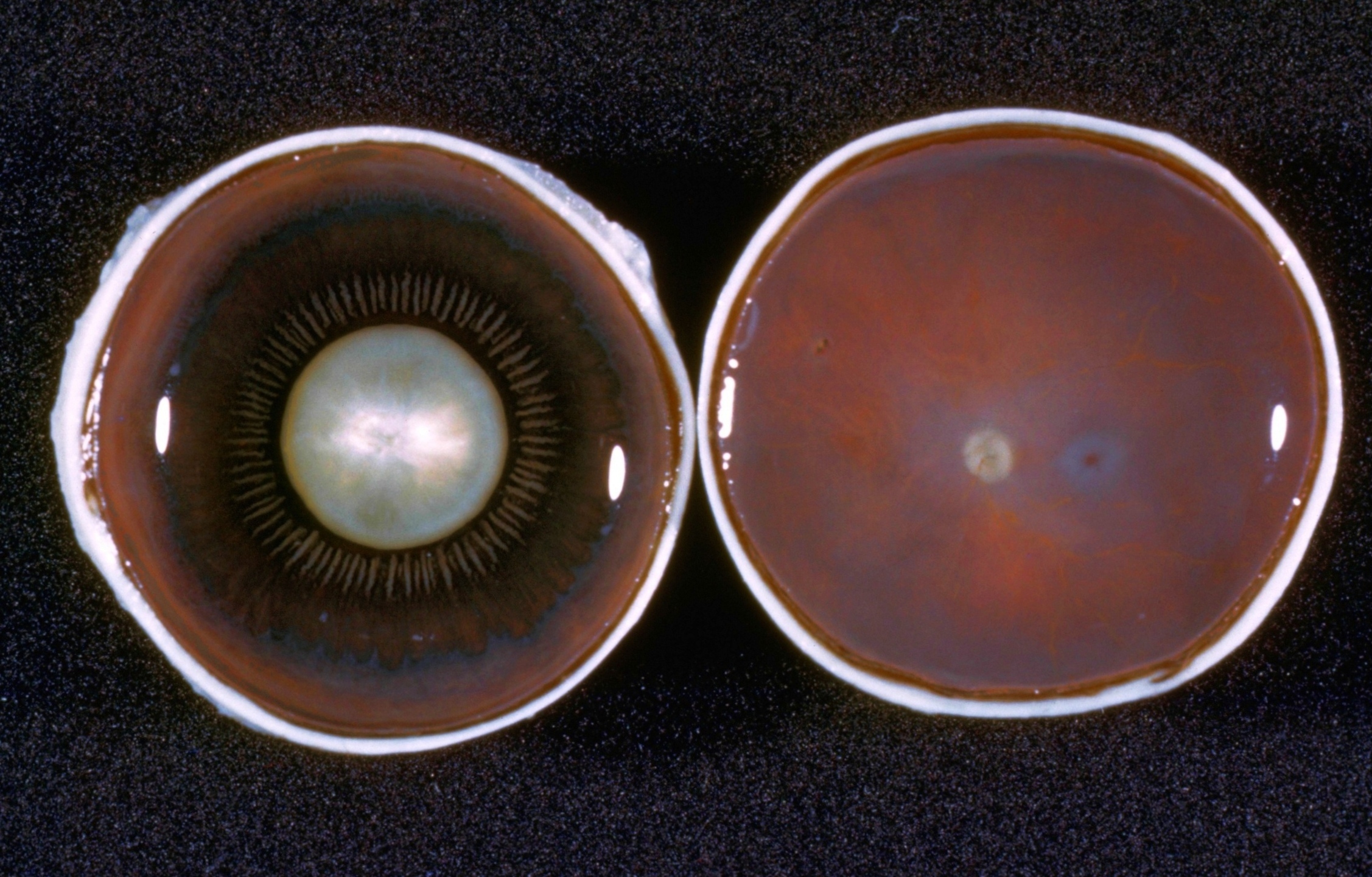
Interior of left eye cut in frontal plane
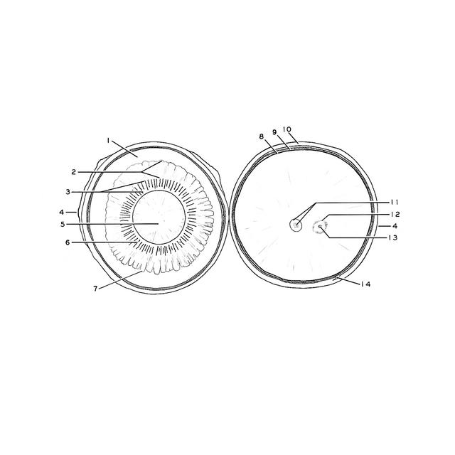
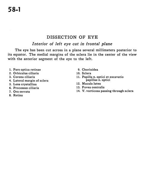
The eye has been cut across in a plane several millimeters posterior to its equator. The medial margins of the sclera lie in the center of the view with the anterior segment of the eye to the left.
- Optic (visual) part of retina
- Orbiculus ciliaris
- Corona ciliaris
- Lateral margin of sclera
- Crystalline lens
- Ciliary process
- Ora serrata
- Retina
- Choroid
- Sclera
- Papilla of optic nerve and hollowed area for papilla
- Macula lutea
- Fovea centralis
- Vorticose vein passing through sclera