For permissions information regarding the use of these images,
please contact Drew Bourn at dbourn@stanford.edu
Image #57-2
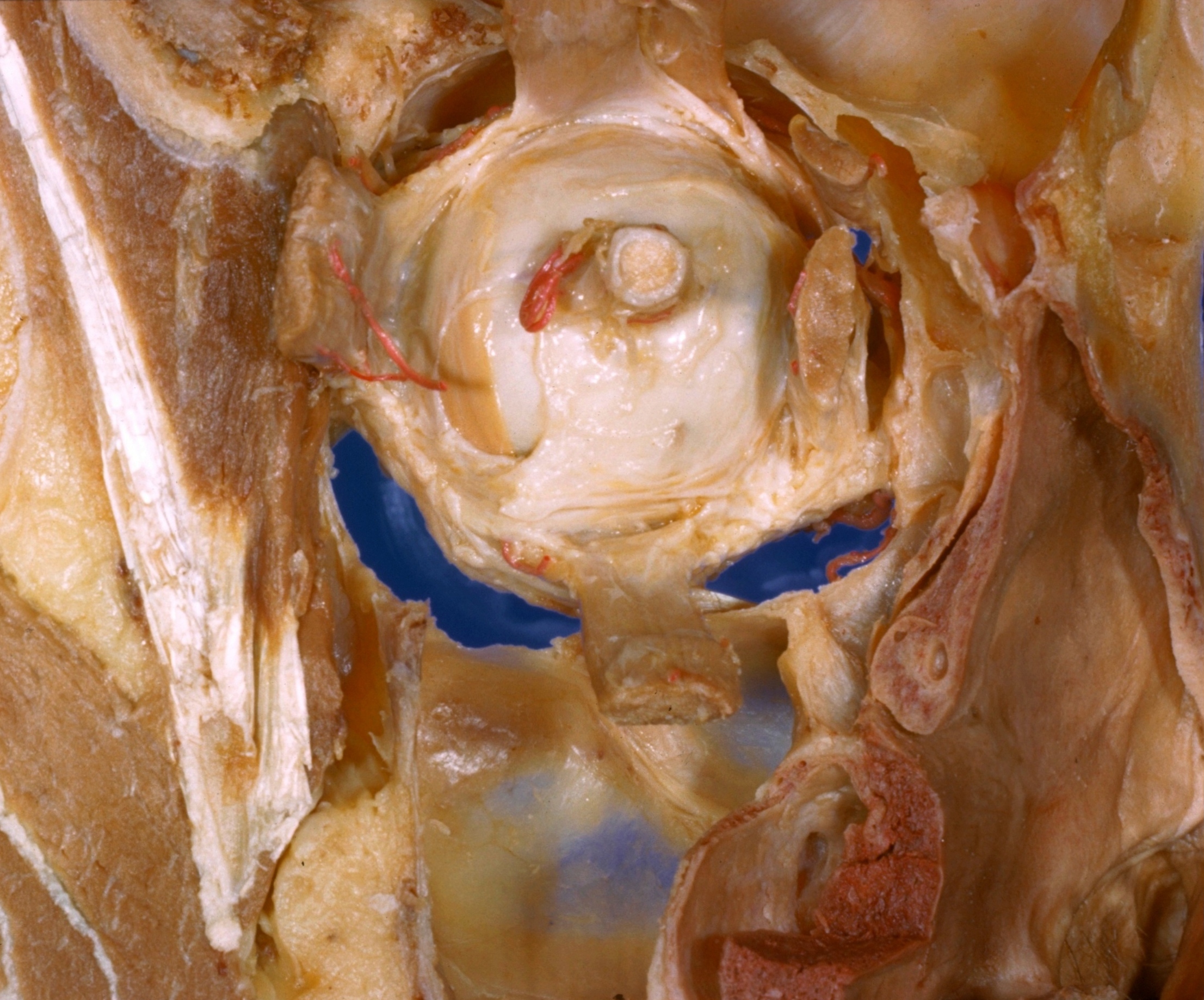
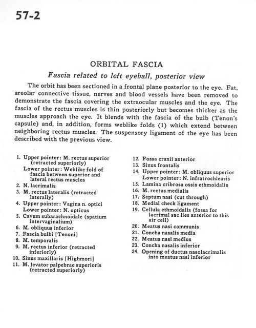
Orbital fascia
Fascia related to left eyeball, posterior view
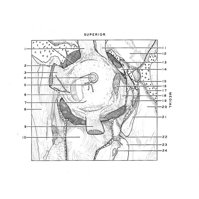
The orbit has been sectioned in a frontal plane posterior to the eye. Fat, areolar connective tissue, nerves and blood vessels have been removed to demonstrate the fascia covering the extraordinary muscles and the eye. The fascia of the rectus muscles is thin posteriorly but becomes thick as the muscles approach the eye. It blends with the fascia of the bulb (Tenon's capsule) and, in addition, forms weblike folds (1) which extend between neighboring rectus muscles. The suspensory ligament of the eye has been described with the previous view.
1
.
Upper pointer: Superior rectus muscle (retracted superiorly) Lower pointer: Weblike fold of fascia between superior and lateral rectus muscles
2
.
Lacrimal nerve
3
.
Lateral rectus muscle (retracted laterally)
4
.
Upper pointer: Sheath of optic nerve Lower pointer: Optic nerve