Dissection of left orbit from a lateral approach
Relation of lacrimal gland to periorbita and zygomatic nerve
Stanford holds the copyright to the David L. Bassett anatomical images and has assigned
Creative Commons license Attribution-Share
Alike 4.0 International to all of the images.
For additional information regarding use and permissions,
please contact Dr. Drew Bourn at dbourn@stanford.edu.
Image #55-2
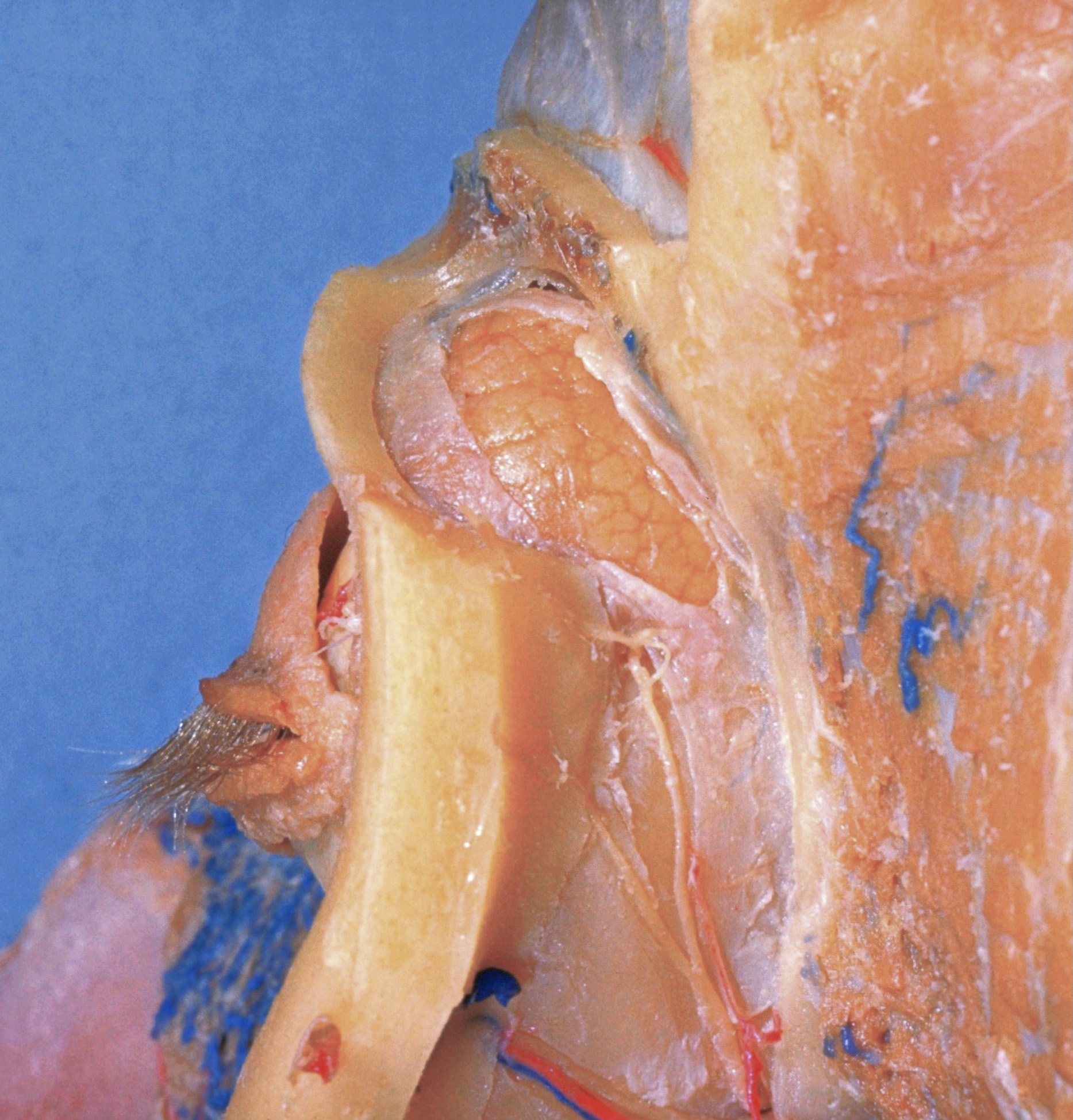
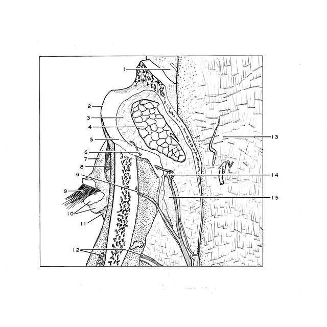
Dissection of left orbit from a lateral approach
Relation of lacrimal gland to periorbita and zygomatic nerve
The roof and lateral wall of the orbit have been cut away. The bony orbital margin has been preserved and the anterior wall of the middle cranial fossa (great wing of sphenoid bone) has been left undisturbed.
- Dura mater within anterior cranial fossa
- Supraorbital margin
- Periorbita (partially cut away to reveal lacrimal gland)
- Superior lacrimal gland
- Zygomaticofrontal suture
- Zygomaticotemporal branches zygomatic nerve
- Superior tarsus
- Lacrimal nerve (terminal branches)
- Lashes
- Upper pointer: Margin of skin Lower pointer: Inferior tarsus
- Conjunctiva
- Zygomaticofacial branches of zygomatic nerve
- Temporal fossa (origin of temporalis muscle)
- Anastomotic branch lacrimal nerve with zygomatic nerve
- Orbital fat pad