Dissection of left orbit from an anterior approach
Orbicularis oculi muscle; superficial nerves and blood vessels
Stanford holds the copyright to the David L. Bassett anatomical images and has assigned
Creative Commons license Attribution-Share
Alike 4.0 International to all of the images.
For additional information regarding use and permissions,
please contact Dr. Drew Bourn at dbourn@stanford.edu.
Image #52-2
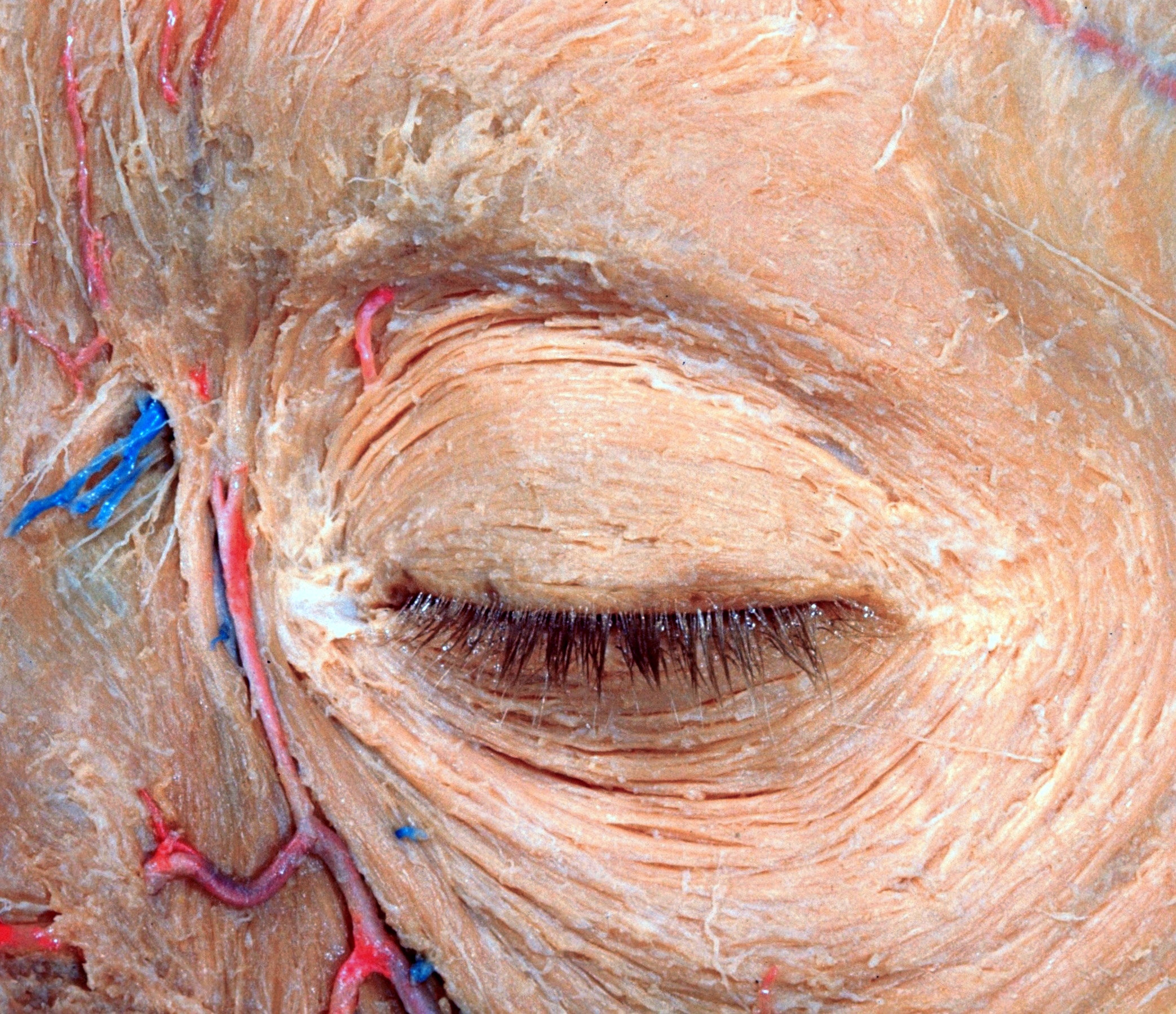
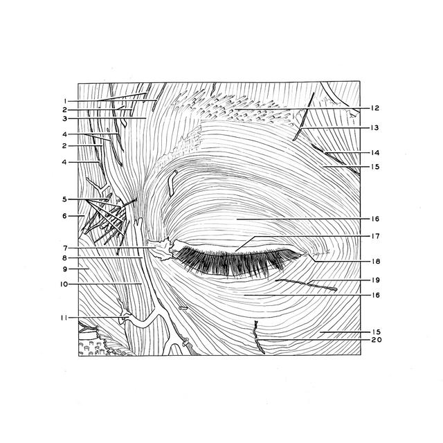
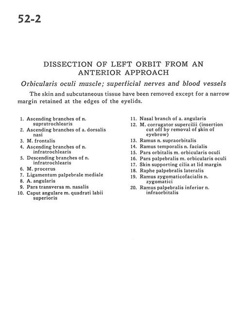
Dissection of left orbit from an anterior approach
Orbicularis oculi muscle; superficial nerves and blood vessels
The skin and subcutaneous tissue have been removed except for a narrow margin retained at the edges of the eyelids.
- Ascending branches of supratrochlear nerve
- Ascending branches of dorsal nasal artery
- Frontalis muscle
- Ascending branches of infratrochlear nerve
- Descending branches of infratrochlear nerve
- Procerus muscle
- Medial palpebral ligament
- Angular artery
- Transverse part of nasalis muscle
- Angular head of levator Iabii superioris muscle
- Nasal branch of angular artery
- Corrugator supercilii muscle (insertion cut off by removal of skin of eyebrow)
- Branch supraorbital nerve
- Temporal branch facial nerve
- Orbital part orbicularis oculi muscle
- Palpebral part of orbicularis oculi muscle
- Skin supporting lashes at lid margin
- Lateral palpebral raphe (ligament)
- Zygomaticofacial branch of zygomatic nerve
- Inferior palpebral branch of infraorbital nerve