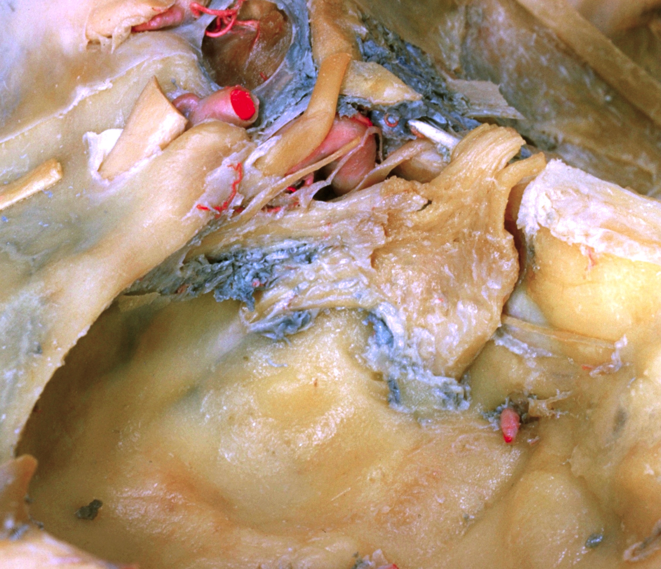
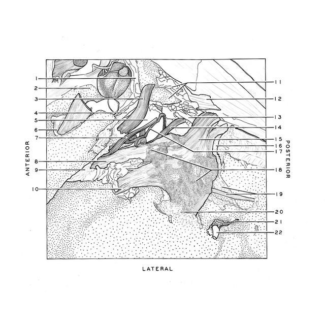
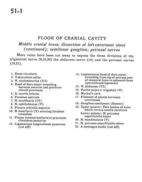
Floor of cranial cavity
Middle cranial fossa; dissection of left cavernous sinus (continued); semilunar ganglion; petrosal nerves
Stanford holds the copyright to the David L. Bassett anatomical images and has assigned
Creative Commons license Attribution-Share Alike 4.0 International to all of the images.
For additional information regarding use and permissions,
please contact Dr. Drew Bourn at dbourn@stanford.edu.
Image #51-1
 |  | ||||||||||||||||||||||||||||||||||||||||||||||||
 |
|