X CLOSE

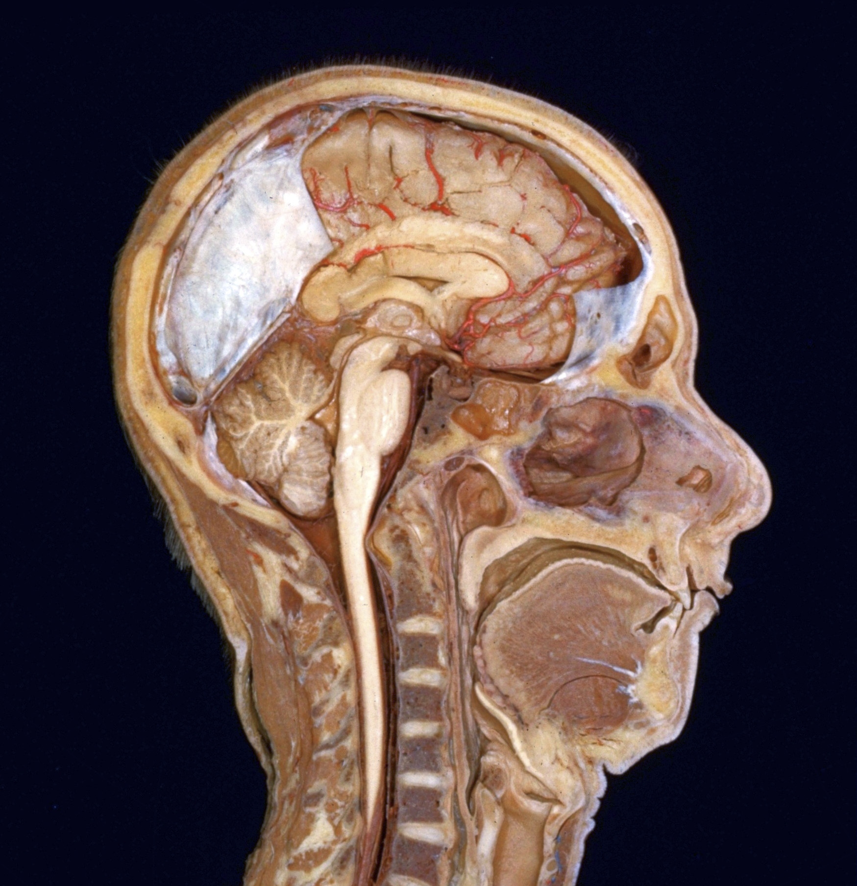
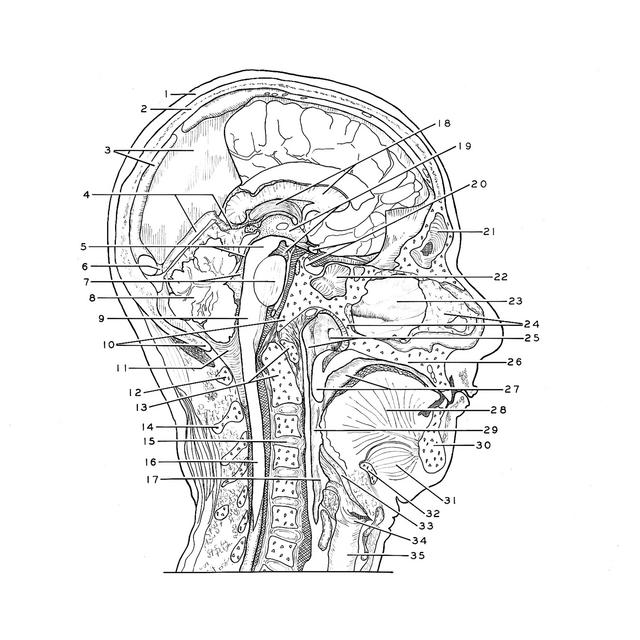
General orientation views of dissection

Midsagittal section of head and neck, right lateral view

For permissions information regarding the use of these images, please contact Drew Bourn at dbourn@stanford.edu

Image #50-1
Bassett ImageDiagram Image
Legend Image
General orientation views of dissection
Midsagittal section of head and neck, right lateral view
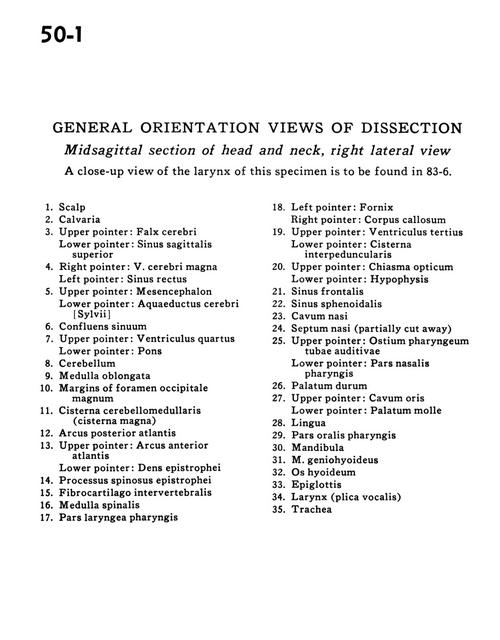
A close-up view of the larynx of this specimen is to be found in 83-6.
1 . Scalp
2 . Calvaria
3 . Upper pointer: Falx cerebri Lower pointer: Superior sagittal sinus
4 . Right pointer: Great cerebal vein Left pointer: Straight sinus
5 . Upper pointer: Mesencephalon Lower pointer: Cerebral aqueduct
6 . Confluence of the sinuses
7 . Upper pointer: Fourth ventricle Lower pointer: Pons
8 . Cerebellum
9 . Medulla oblongata
10 . Margins of foramen magnum
11 . Cerebellomedullary cistern (cisterna magna)
12 . Posterior arch atlas
13 . Upper pointer: Anterior arch atlas Lower pointer: Dens axis
14 . Spinous process axis
15 . Intervertebral disc
16 . Spinal cord
17 . Laryngeal part of pharynx
18 . Left pointer: Fornix Right pointer: Corpus callosum
19 . Upper pointer: Third ventricle Lower pointer: Interpeduncular cistern
20 . Upper pointer: Optic chiasm Lower pointer: Hypophysis
21 . Frontal sinus
22 . Sphenoid sinus
23 . Nasal cavity
24 . Nasal septum (partially cut away)
25 . Upper pointer: Bony auditory tube Lower pointer: Nasal part pharynx
26 . Hard palate
27 . Upper pointer: Oral cavity Lower pointer: Soft palate
28 . Tongue
29 . Oral part pharynx
30 . Mandible
31 . Geniohyoid muscle
32 . Hyoid bone
33 . Epiglottis
34 . Larynx (plica vocalis)
35 . Trachea