Deep facial dissection, lateral view; temporal muscle; course of facial nerve through parotid gland
For permissions information regarding the use of these images,
please contact Drew Bourn at dbourn@stanford.edu
Image #49-5
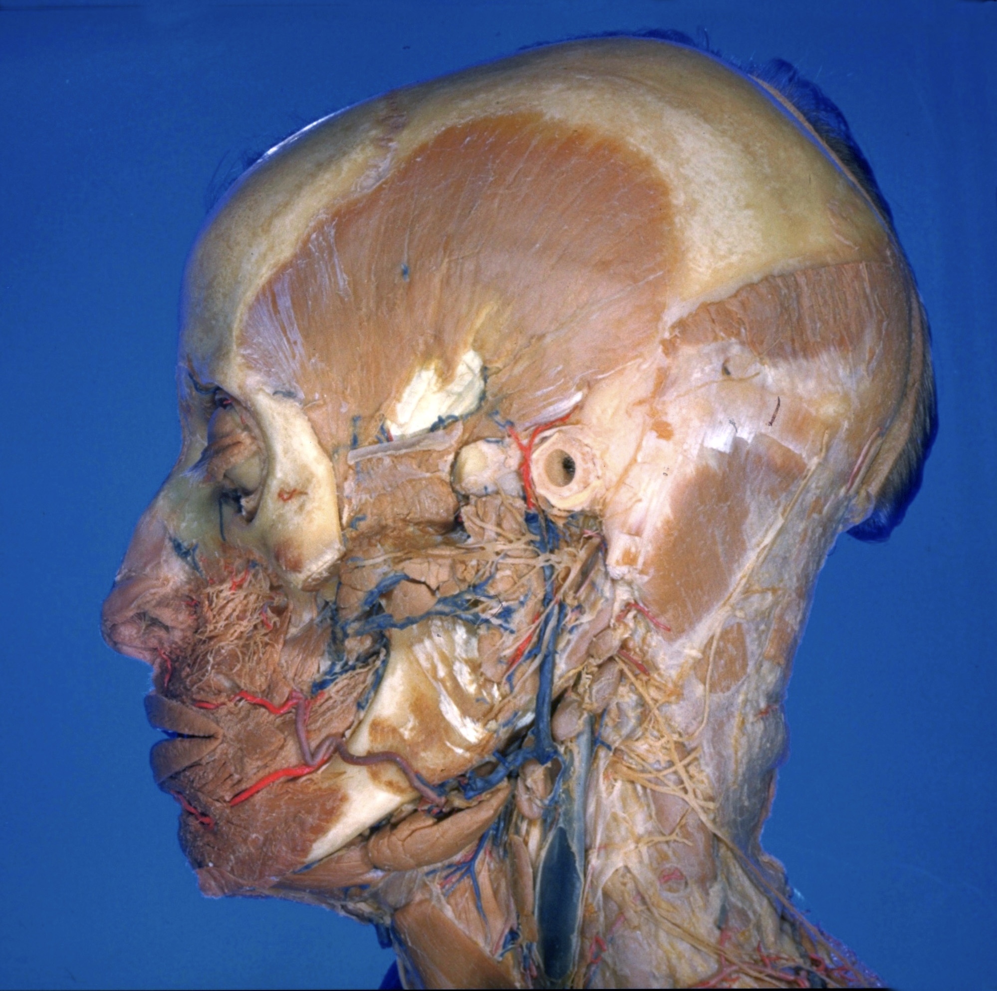
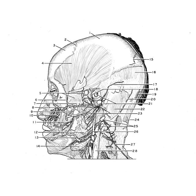
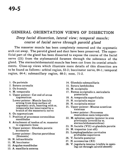
General orientation views of dissection
Deep facial dissection, lateral view; temporal muscle; course of facial nerve through parotid gland
The masseter muscle has been completely removed and the zygomatic arch cut away. The parotid gland and duct have been preserved. The superficial part of the gland has been dissected to expose the course of the facial nerve (23) from the stylomastoid foramen through the substance of the gland. The sternocleidomastoid muscle has been cut from its cranial attachments. Close-up views which illustrate more details of this dissection are to be found as follows
1
.
Parietal bone
2
.
Coronal suture
3
.
Frontal bone
4
.
Temporalis muscle
5
.
Upper pointer: Cut end of zygomatic arch Lower pointer: Muscle tendon arising from deep surface of zygomatic arch, inserting with temporalis muscle but innervated by a branch of the masseteric nerve
6
.
Zygomatic bone
7
.
Position of coronoid process of mandible
8
.
Attachment of tendon of masseter muscle to zygomatic bone