X CLOSE

Osteology

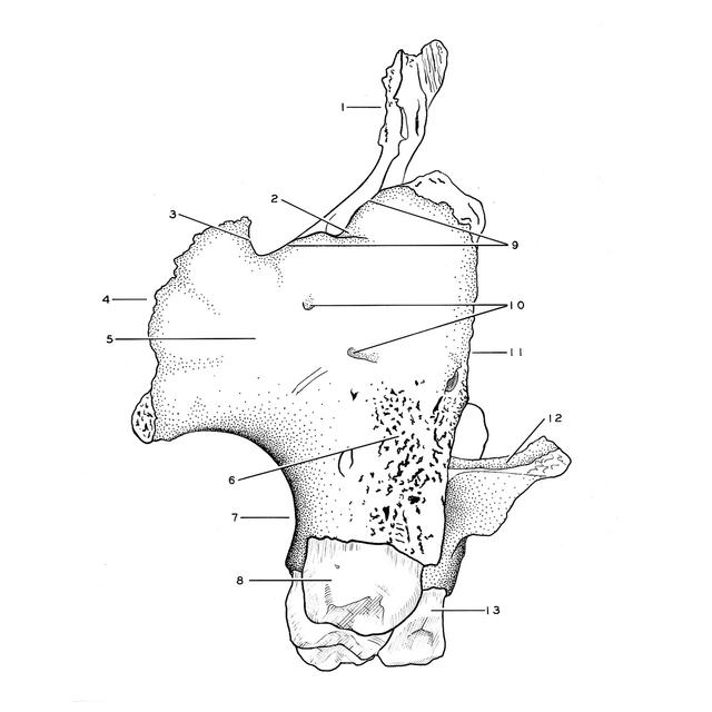
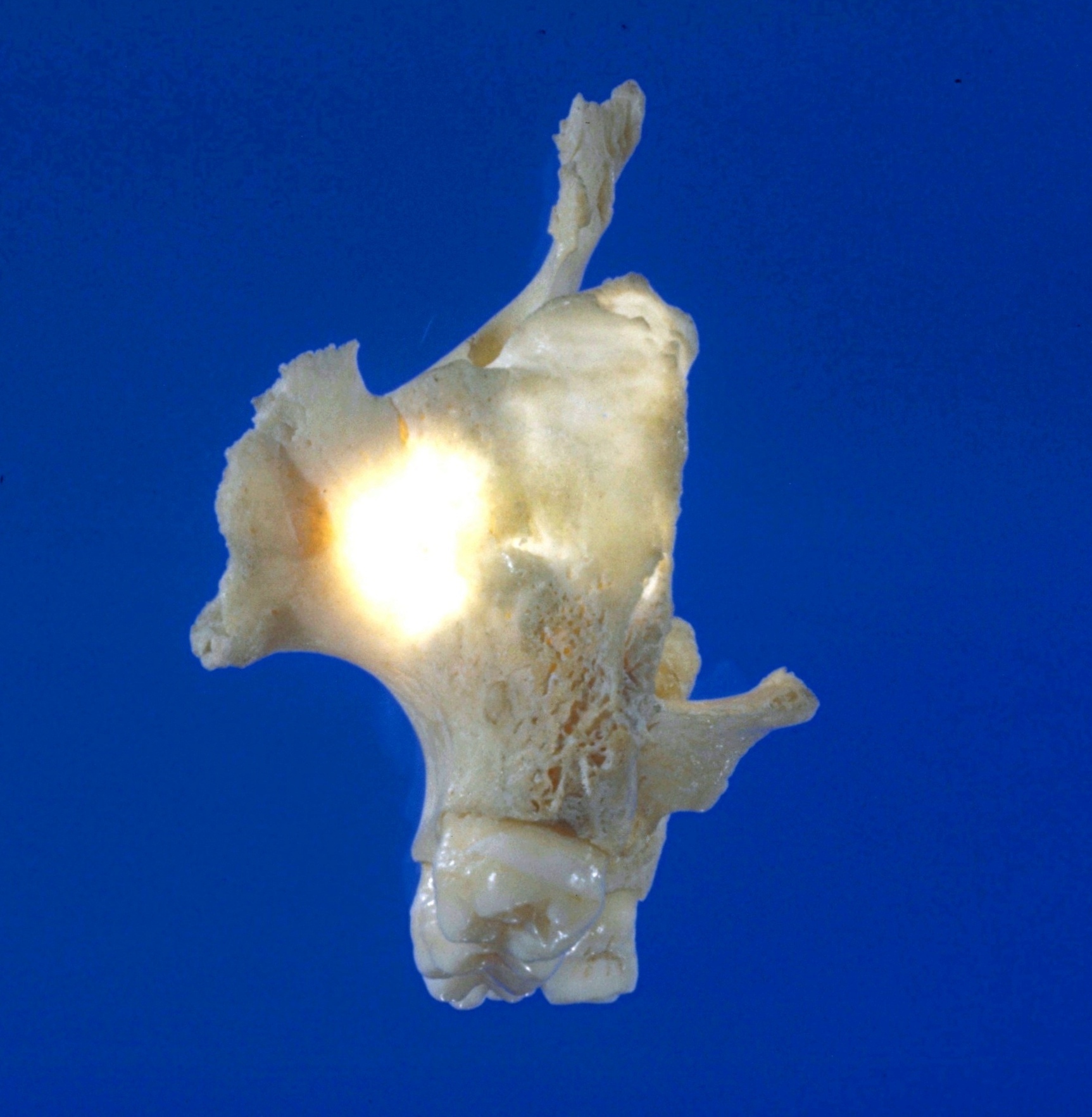
Left maxilla, posterior view

Stanford holds the copyright to the David L. Bassett anatomical images and has assigned Creative Commons license Attribution-Share Alike 4.0 International to all of the images.
For additional information regarding use and permissions, please contact Dr. Drew Bourn at dbourn@stanford.edu.

Image #45-5
Bassett Image
Diagram Image
Legend Image
Osteology
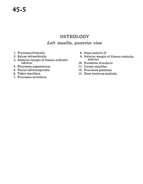
Left maxilla, posterior view
  1. Frontal process
  2. Infraorbital sulcus
  3. Anterior margin of inferior orbital fissure
  4. Zygomatic process
  5. Infratemporal surface
  6. Maxillary tubercle
  7. Alveolar process
  8. Molar II
  9. Inferior margin of inferior orbital fissure
  10. Alveolar foramen
  11. Body maxilla
  12. Palatine process
  13. Medial incisor