Osteology
Roentgenogram, ethmoid bone, anteroposterior view
Stanford holds the copyright to the David L. Bassett anatomical images and has assigned
Creative Commons license Attribution-Share
Alike 4.0 International to all of the images.
For additional information regarding use and permissions,
please contact Dr. Drew Bourn at dbourn@stanford.edu.
Image #43-4
Osteology
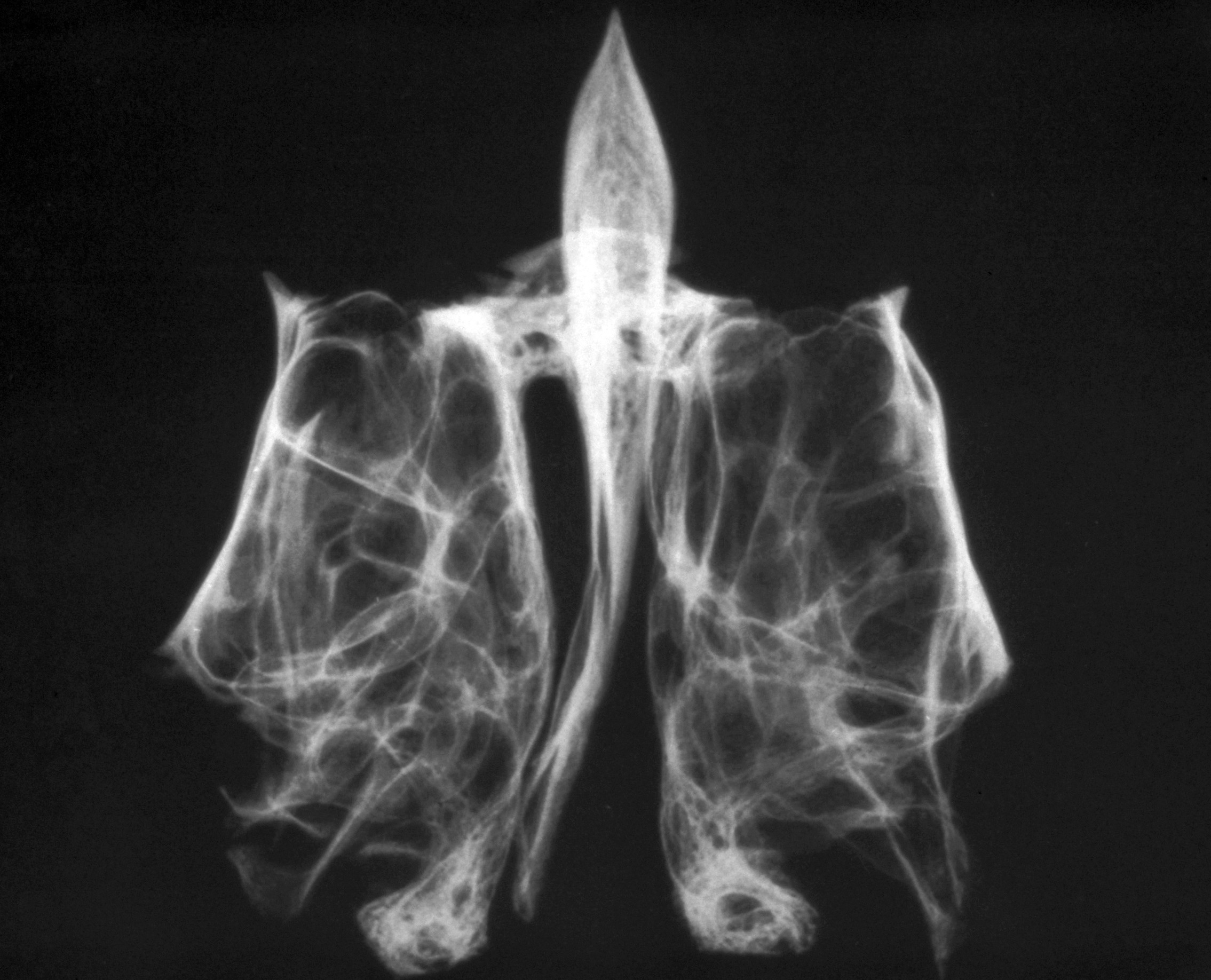
Roentgenogram, ethmoid bone, anteroposterior view
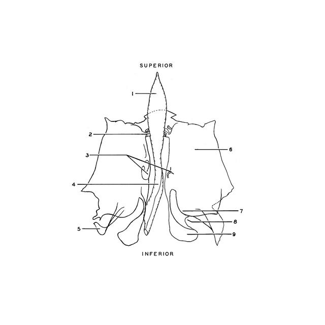
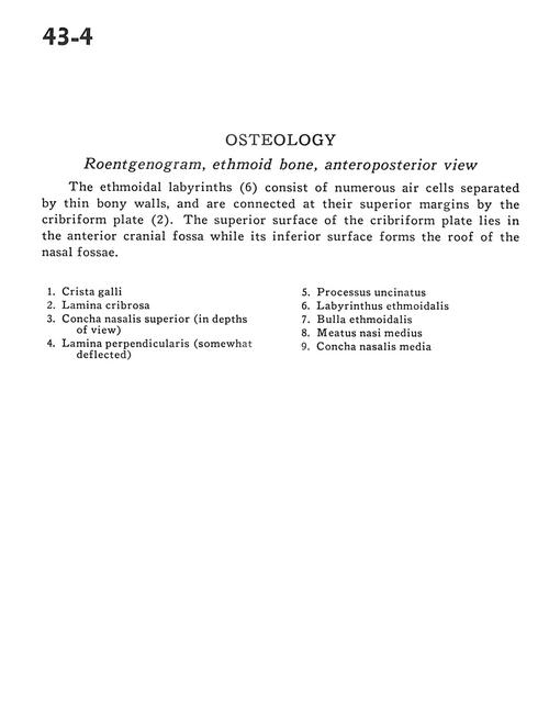
The ethmoidal labyrinths (6) consist of numerous air cells separated by thin bony walls, and are connected at their superior margins by the cribriform plate (2). The superior surface of the cribriform plate lies in the anterior cranial fossa while its inferior surface forms the roof of the nasal fossae.
- Crista galli
- Crista galli
- Alar process
- Cribriform plate
- Sulci for anterior ethmoidal vessels and nerves
- Superior nasal concha (in depths of view)
- Cribriform plate
- Perpendicular plate (somewhat deflected)
- Lamina papyracea
- Uncinate process
- Uncinate process
- Ethmoidal labyrinth
- Middle nasal concha
- Ethmoidal bulla
- Ethmoidal labyrinth
- Middle nasal meatus
- Articular border for frontal spine
- Middle nasal concha
- Ethmoidal cells
- Perpendicular plate
- Middle nasal meatus