For permissions information regarding the use of these images,
please contact Drew Bourn at dbourn@stanford.edu
Image #38-2
Osteology
Lateral wall of left nasal fossa dissected
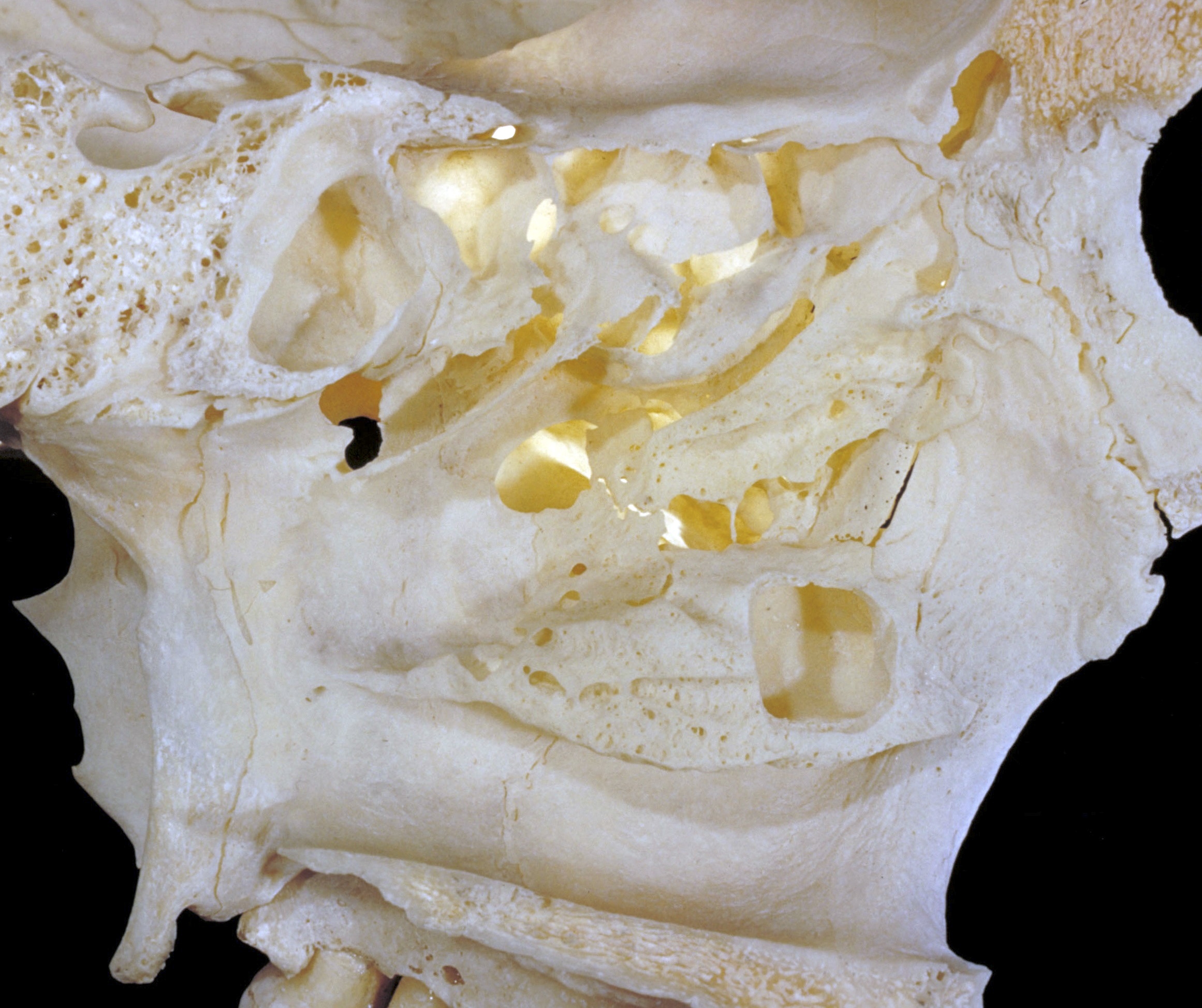
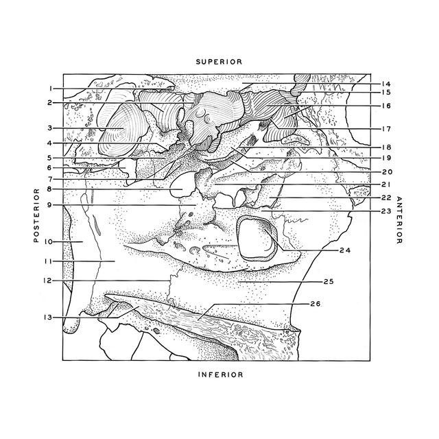
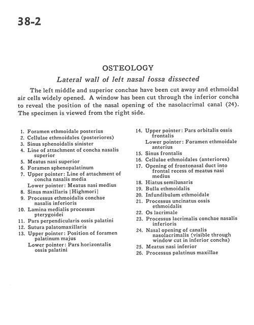
The left middle and superior conchae have been cut away and ethmoidal air cells widely opened. A window has been cut through the inferior concha to reveal the position of the nasal opening of the nasolacrimal canal (24). The specimen is viewed from the right side.
1
.
Posterior ethmoidal foramen
2
.
Ethmoidal cells (posterior)
3
.
Sphenoid sinus left
4
.
Line of attachment of superior nasal concha
5
.
Superior nasal meatus
6
.
Sphenopalatine foramen
7
.
Upper pointer: Line of attachment of middle nasal concha Lower pointer: Middle nasal meatus
8
.
Maxillary sinus
9
.
Ethmoidal process inferior nasal conchae
10
.
Medial plate of pterygoid process
11
.
Perpendicular part palatine bone
12
.
Palatomaxillary suture
13
.
Upper pointer: Position of greater palatine foramen Lower pointer: Horizontal plate palatine bone
14
.
Upper pointer: Orbital part of frontal bone Lower pointer: Anterior ethmoidal foramen
15
.
Frontal sinus
16
.
Ethmoidal cells (anterior)
17
.
Opening of frontonasal duct into frontal recess of middle nasal meatus
18
.
Hiatus semilunaris
19
.
Ethmoidal bulla
20
.
Ethmoidal infundibulum
21
.
Uncinate process ethmoid bone
22
.
Lacrimal bone
23
.
Lacrimal process inferior nasal conchae
24
.
Nasal opening of nasolacrimal canal visible through window cut in inferior concha)