For permissions information regarding the use of these images,
please contact Drew Bourn at dbourn@stanford.edu
Image #38-1
Osteology
Lateral wall of right nasal fossa
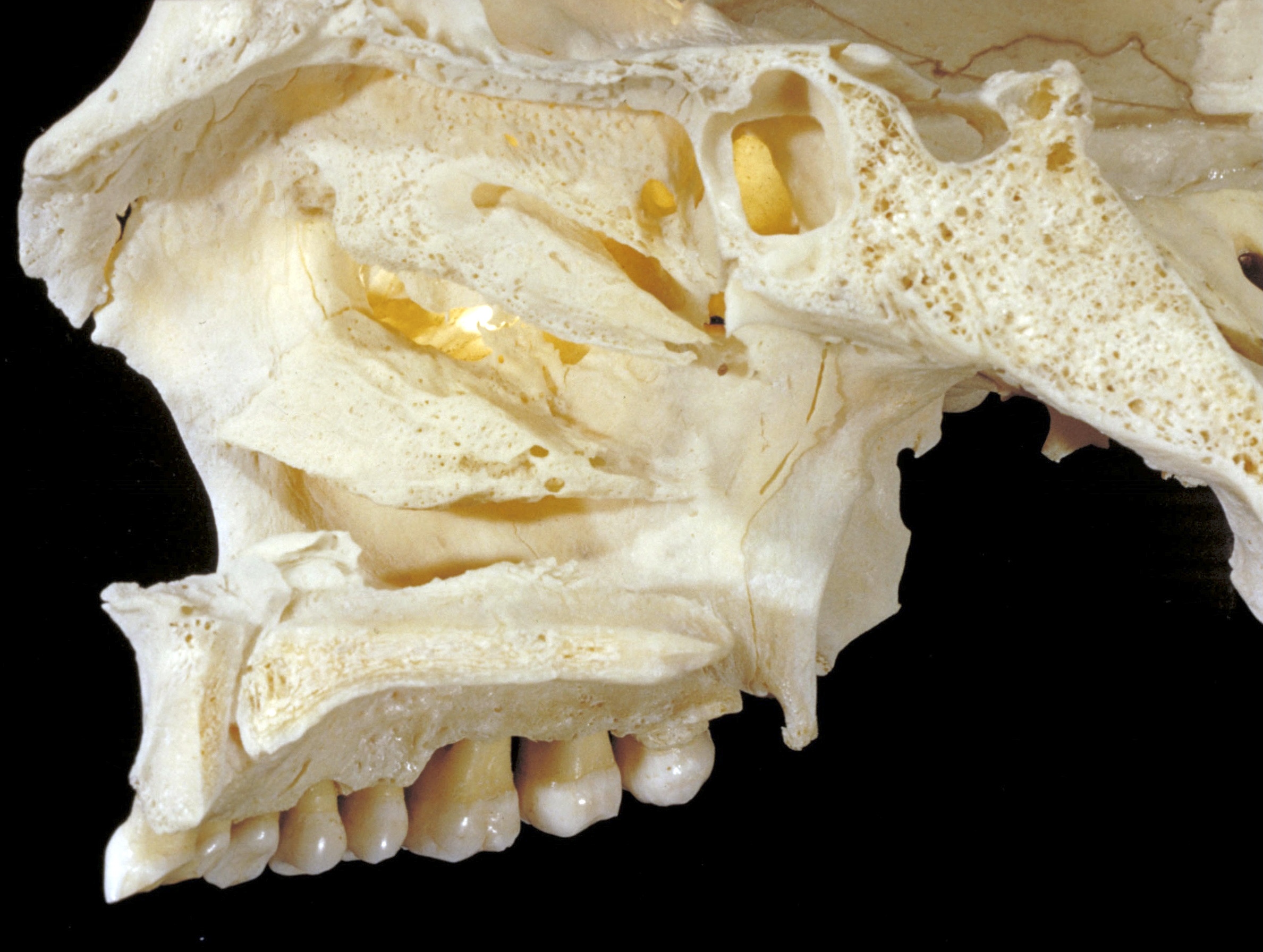
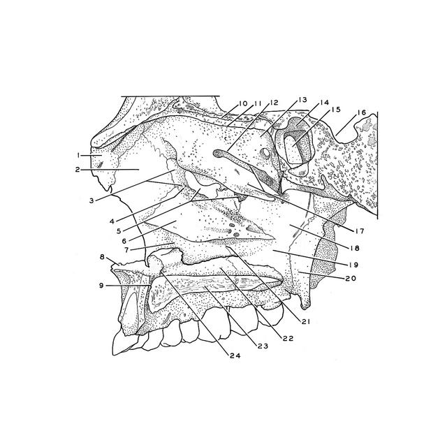
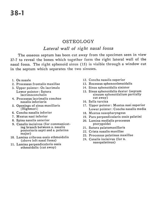
The osseous septum has been cut away from the specimen seen in view 37-7 to reveal the bones which together form the right lateral wall of the nasal fossa. The right sphenoid sinus (15) is visible through a window cut in the septum which separates the two sinuses.
1
.
Nasal bone
2
.
Frontal process of maxilla
3
.
Upper pointer: Lacrimal bone Lower pointer: Lacrimoconchal suture
4
.
Lacrimal process inferior nasal conchae
5
.
Openings of maxillary sinus
6
.
Inferior nasal concha
7
.
Inferior nasal meatus
8
.
Anterior nasal spine
9
.
Incisive canal (for communicating branch between posterior nasal septal artery and greater palatine artery)
10
.
Cribriform plate éthmoid bone (above left nasal fossa)
11
.
Perpendicular plate of ethmoid bone (cut away)
12
.
Superior nasal concha
13
.
Sphenoethmoidal recess
14
.
Sphenoid sinus left
15
.
Sphenoid sinus right (sphenoidal septal sinuses partially cut away)
16
.
Sella turcica
17
.
Upper pointer: Superior nasal meatus Lower pointer: Middle nasal concha