Radiographs of the brain
Postero-anterior (P-A) pneumoencephalogram
Stanford holds the copyright to the David L. Bassett anatomical images and has assigned
Creative Commons license Attribution-Share
Alike 4.0 International to all of the images.
For additional information regarding use and permissions,
please contact Dr. Drew Bourn at dbourn@stanford.edu.
Image #34-7
Radiographs of the brain
Postero-anterior (P-A) pneumoencephalogram
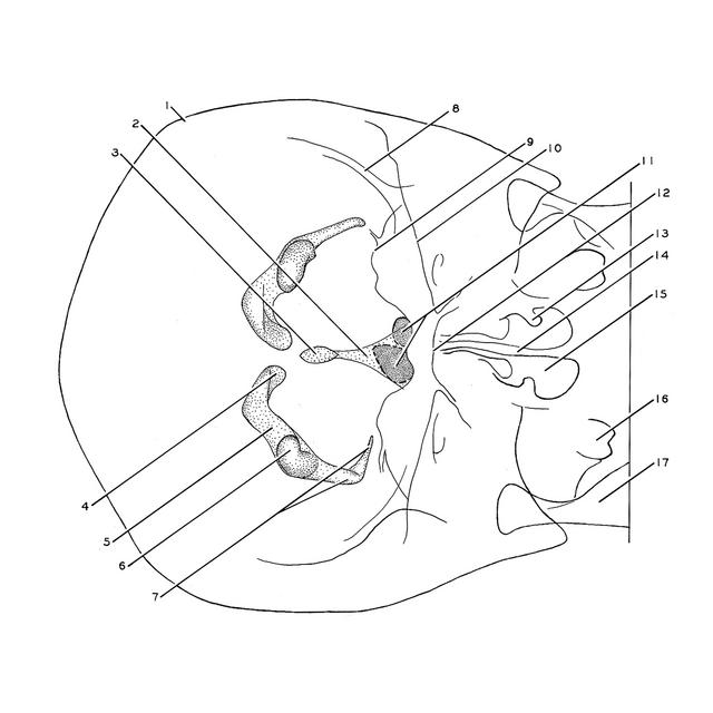
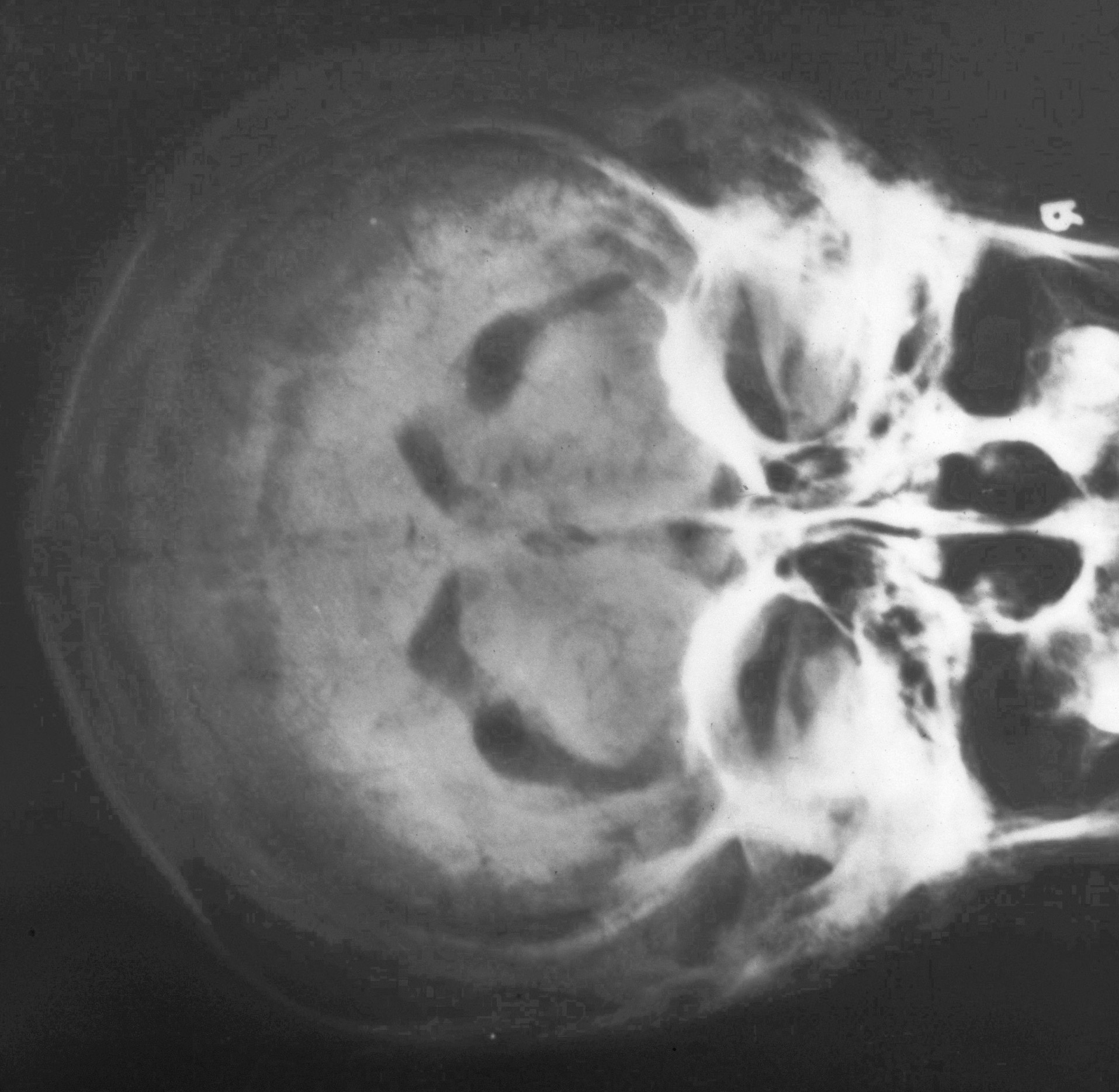
The subject has been placed in a prone position. The lateral ventricles are completely outlined by air. Some air is also contained within the third ventricle (3). The fourth ventricle (2) and the cerebral aqueduct are less distinct and are in part obscured by air in the frontal sinuses (11). Small collections of air (not drawn) can be seen within the subarachnoid spaces in many sulci of the brain. (These films have been reproduced through the courtesy of Dr. Howard L. Jones of the Palo Alto Hospital.)
- Calvaria
- Fourth ventricle
- Third ventricle (partially filled)
- Anterior horn lateral ventricle
- Central part lateral ventricle
- Posterior horn lateral ventricle
- Inferior horn of lateral ventricle
- Temporal surface of frontal bone
- Orbital part of frontal bone
- Outline of petrosal part of temporal bone
- Frontal sinus
- Outline of Basilar part of occipital bone
- Inferior nasal concha
- Bony nasal septum
- Nasal cavity
- Dens
- Ramus of mandible.