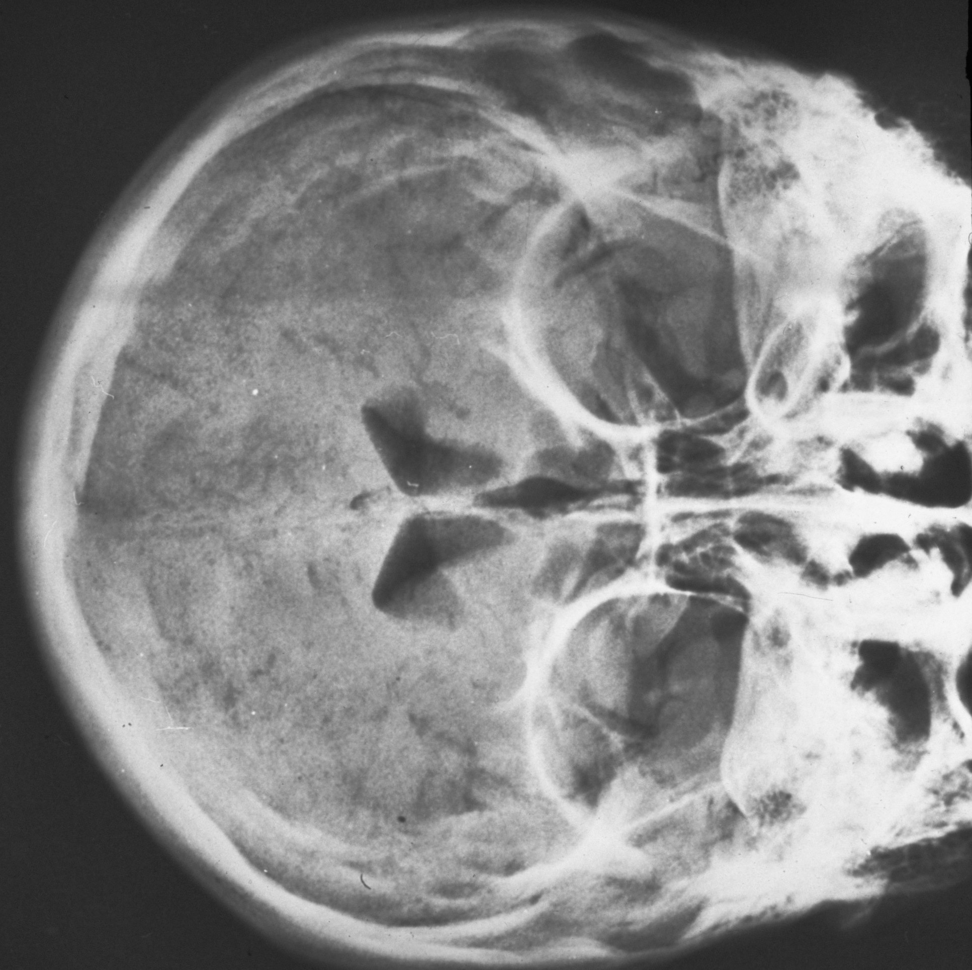
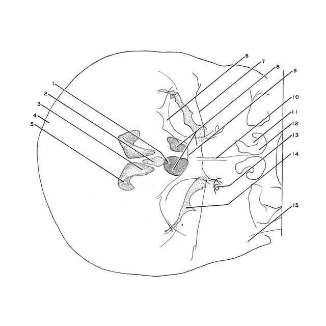
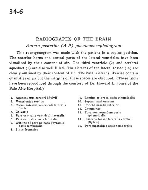
Radiographs of the brain
Antero-posterior (A-P) pneumoencephalogram
Stanford holds the copyright to the David L. Bassett anatomical images and has assigned
Creative Commons license Attribution-Share Alike 4.0 International to all of the images.
For additional information regarding use and permissions,
please contact Dr. Drew Bourn at dbourn@stanford.edu.
Image #34-6
 |  | ||||||||||||||||||||||||||||||||||
 |
|